. 2019 Mar 29;14(3):e0212659.
doi: 10.1371/journal.pone.0212659. eCollection 2019.

Thyroxine (T4) may promote re-epithelialisation and angiogenesis in wounded human skin ex vivo

Affiliations

Thyroxine (T4) may promote re-epithelialisation and angiogenesis in wounded human skin ex vivo

Guo-You Zhang et al. PLoS One. .

Abstract

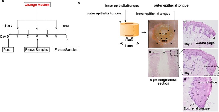
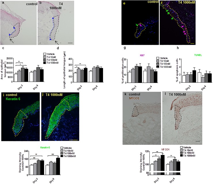
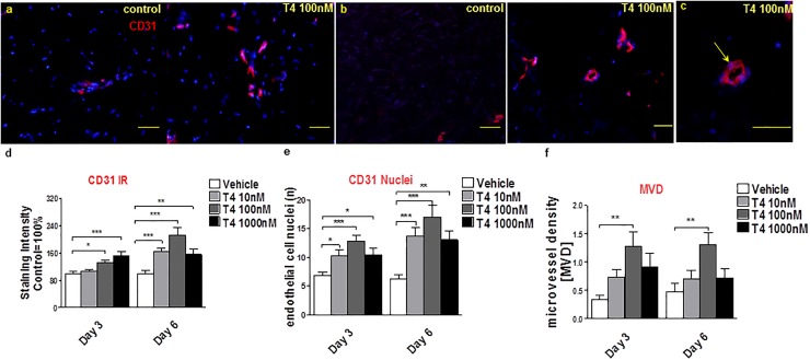
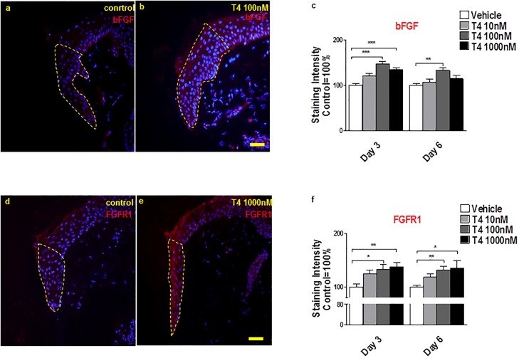
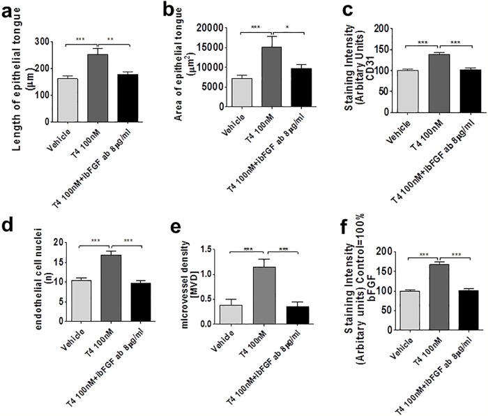
There is a pressing need for improved preclinical model systems in which to study human skin wound healing. Here, we report the development and application of a serum-free full thickness human skin wound healing model. Not only can re-epithelialization (epidermal repair) and angiogenesis be studied in this simple and instructive model, but the model can also be used to identify clinically relevant wound-healing promoting agents, and to dissect underlying candidate mechanisms of action in the target tissue. We present preliminary ex vivo data to suggest that Thyroxine (T4), which reportedly promotes skin wound healing in rodents in vivo, may promote key features of human skin wound healing. Namely, T4 stimulates re-epithelialisation and angiogenesis, and modulates both wound healing-associated epidermal keratin expression and energy metabolism in experimentally wound human skin. Functionally, the wound healing-promoting effects of T4 are at least partially mediated via fibroblast growth factor/fibroblast growth factor receptor-mediated signalling, since they could be significantly antagonized by bFGF-neutralizing antibody. Thus, this pragmatic, easy-to-use full-thickness human skin wound healing model provides a useful preclinical research tool in the search for clinically relevant candidate wound healing-promoting agents. These ex vivo data encourage further pre-clinical testing of topical T4 as a cost-efficient, novel agent in the management of chronic human skin wounds.


Conflict of interest statement

The authors have declared that no competing interests exist.

Figures

Fig 1
Fig 1. Establishment of human wound healing assay and testing of effects of T4 on keratinocyte migration during epidermal repair of experimentally wound human skin.
(a) A schematic diagram of human wound healing culture model assay. A schematic diagram (b) and example of a wound punch sample (c) and longitudinal section (d). Sections from day 0 and day 6 of culture (vehicle control) are shown (e-g). The regenerative phenomenon is highlighted in (f). Magnification of marked area in (f) is shown in (g).
Fig 2
Fig 2. T4 promotes human skin re-epithelialisation.
(a-d) Haematoxylin and eosin histochemistry: overview of wounded human skin fragment. After 3 days, epithelial tongue areas (blacked dotted line area) (ET) and length (blue dotted line) were significantly greater after treatment with T4 compared to control alone. (e-h) Cryosections of control-, or T4-treated human skin were examined by Ki-67 (red arrow)/TUNEL (green arrow) double-labelling [19, 23, 24, 56, 57] (e, f). The percentage of positive cells was analyzed in the new ETs (see dotted line). When compared to the control group, more Ki-67 positive cells after 3 day culture with 1000 nM T4 treatment (and more TUNEL positive cells at day 3 in T4 1000 nM treated group were found, although the differences were not statistically significant (e-h). (i, j) CK6 expression was significantly upregulated by T4, especially in the 1000 nM treatment group. Green fluorescence staining represented cytokeratin 6 IR in the new wound ET (dotted line area).(k, l) MTCO1 expression was significantly up-regulated by T4. Brown staining represents MTCO1 IR in the new ET. Staining intensity was measured in a defined reference area (dotted line) and normalized to the control (100%), as for CK6 expression. Number of independent experiments: n = 3 subjects (i.e. 1–2 punches per patient, per treatment group and per time point and at least 8 photomicrographs were analysed per condition); data were pooled since the results trends in all three independent experiments were comparable). *p<0.05, **p<0.01, ***<0.001. Scale bars = 50μm.
Fig 3
Fig 3. T4 stimulates angiogenesis in wounded human skin.
(a-c) To analyze angiogenesis, the number of CD31+ cells (red) and of CD31+ blood vessel cross-sections (lumina) (yellow arrow, c) per visual field were counted by immunofluorescence microscopy (at least 12 visual fields per skin fragment were evaluated). In addition, the intensity of CD31 IR was measured. Scale bars in a, b = 50μm, c = 200μm. (d) CD31 IR was significantly up-regulated by T4 at days 3 and 6. Immunoreactivity data was normalized to the control data as were (e, f) the number of CD31 +ve endothelial cell nuclei (CD31+/DAPI+ cells) and lumina per microscopic field. Number of independent experiments: n = 3 subjects (i.e. 1–2 punches per patient, per treatment group and per time point and at least 8 photomicrographs were analyzed per condition); data were pooled since the results trends in all three independent experiments were comparable). MVD: Microvessel density; ibFGF ab: inhibitory bFGF antibody.
Fig 4
Fig 4. T4 up-regulates bFGF and FGF receptor 1 IR.
(a-c) bFGF expression was significantly up-regulated by T4 in the epidermis of new epithelial tongues (staining intensity was measured in reference area) at day 3 and day 6 in 100nM T4 condition. Red fluorescence staining represents bFGF IR in the new epithelial tongue and the control IR value was normalized as 100%. (d-f) T4 increases FGFR1 expression. Red fluorescence staining represents FGFR1 IR in the new epithelial tongue. Staining intensity was measured in the dotted line reference area and the control IR value was normalized as 100%. One-Way ANOVA by appropriate post hoc comparisons was used. Data represent the mean±SEM of 3 independent experiments and normalized to the control as 100%. 1–2 punches per patient, per treatment group and per time point and at least 8 photomicrographs were analyzed per condition. *P<0.05; **P<0.01; ***P<0.001. Scale bars = 50μm.
Fig 5
Fig 5. (a-f) T4 mediates its effect at least partially via bFGF.
T4 exerts its effect partly through inducing bFGF at day 3. Area and length of the ET (a: length of new ET; b: area of new ET), intensity of the IR of CD31 (c), number of CD31+ cells (d), Number of CD31+ lumina (e), and intensity of IR of bFGF in new ET (f) were significantly increased in the T4-treated test group compared to controls. 1–2 punches per patient, per treatment group and per time point and at least 8 photomicrographs were analyzed per condition. These effects could be abrogated by co-treatment with inhibitory bFGF antibody [45].

References

    1. Ilonzo N, Patel M, Lantis JC 2nd. Managing the Diabetic Foot Ulcer: How Best Practices Fit the Real 2018 United States. Surg Technol Int. 2018;32:49–59. Epub 2018/04/04. . - PubMed
    1. Guest JF, Fuller GW, Vowden P. Venous leg ulcer management in clinical practice in the UK: costs and outcomes. Int Wound J. 2018;15(1):29–37. Epub 2017/12/16. 10.1111/iwj.12814 . - DOI - PMC - PubMed
    1. Coleman S, Smith IL, McGinnis E, Keen J, Muir D, Wilson L, et al. Clinical evaluation of a new pressure ulcer risk assessment instrument, the Pressure Ulcer Risk Primary or Secondary Evaluation Tool (PURPOSE T). J Adv Nurs. 2018;74(2):407–24. Epub 2017/08/24. 10.1111/jan.13444 . - DOI - PMC - PubMed
    1. Xie T, Ye J, Rerkasem K, Mani R. The venous ulcer continues to be a clinical challenge: an update. Burns Trauma. 2018;6:18 Epub 2018/06/27. 10.1186/s41038-018-0119-y . - DOI - PMC - PubMed
    1. Jeffcoate WJ, Vileikyte L, Boyko EJ, Armstrong DG, Boulton AJM. Current Challenges and Opportunities in the Prevention and Management of Diabetic Foot Ulcers. Diabetes Care. 2018;41(4):645–52. Epub 2018/03/22. 10.2337/dc17-1836 . - DOI - PubMed

Publication types