. 2019 Jun:195:61-70.
doi: 10.1016/j.jinorgbio.2019.03.009. Epub 2019 Mar 21.

The hydrogen bonding network of coproheme in coproheme decarboxylase from Listeria monocytogenes: Effect on structure and catalysis

Affiliations

The hydrogen bonding network of coproheme in coproheme decarboxylase from Listeria monocytogenes: Effect on structure and catalysis

Lisa Milazzo et al. J Inorg Biochem. 2019 Jun.

Abstract

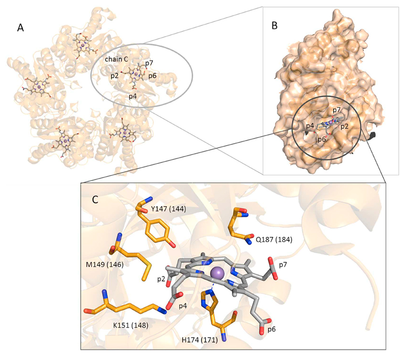
Coproheme decarboxylase (ChdC) catalyzes the oxidative decarboxylation of coproheme to heme b, i.e. the last step in the recently described coproporphyrin-dependent pathway. Coproheme decarboxylation from Listeria monocytogenes is a robust enzymatic reaction of low catalytic efficiency. Coproheme acts as both substrate and redox cofactor activated by H2O2. It fully depends on the catalytic Y147 close to the propionyl group at position 2. In the present study we have investigated the effect of disruption of the comprehensive and conserved hydrogen bonding network between the four propionates and heme cavity residues on (i) the conformational stability of the heme cavity, (ii) the electronic configuration of the ferric redox cofactor/substrate, (iii) the binding of carbon monoxide and, (iv) the decarboxylation reaction mediated by addition of H2O2. Nine single, double and triple mutants of ChdC from Listeria monocytogenes were produced in E. coli. The respective coproheme- and heme b-complexed proteins were studied by UV-Vis, resonance Raman, circular dichroism spectroscopy, and mass spectrometry. Interactions of propionates 2 and 4 with residues in the hydrophobic cavity are crucial for maintenance of the heme cavity architecture, for the mobile distal glutamine to interact with carbon monoxide, and to keep the heme cavity in a closed conformation during turnover. By contrast, the impact of substitution of residues interacting with solvent exposed propionates 6 and 7 was negligible. Except for Y147A and K151A all mutant ChdCs exhibited a wild-type-like catalytic activity. The findings are discussed with respect to the structure-function relationships of ChdCs.

Keywords: Carbon monoxide; Coproheme decarboxylase; Heme b biosynthesis; Propionyl hydrogen-bond; Resonance Raman spectroscopy.


Figures

Fig. 1
Fig. 1
Structure of coproheme decarboxylase (ChdC). (A) Overall structure of coproheme decarboxylase from Geobacillus stearothermophilus (GsChdC) in complex with Mn-coproporphyrin III (pdb-code 5T2K) [9]. (B) Representation of chain C of GsChdC showing solvent exposed propionates p6 and p7. (C) Presentation of the active site of GsChdC with residue numbering for coproheme decarboxylase from Listeria monocytogenes (LmChdC) (GsChdC numbering is given in brackets). Fully conserved residues include the catalytic tyrosine 147, methionine 149, lysine 151, mobile distal glutamine 187 and the proximal histidine 174.
Fig. 2
Fig. 2
Hydrogen-bonding network involving the four coproheme propionate groups of Mn-coproheme-GsChdC (chain C, 5T2K) [9]. (A) Propionates at positions 2 (p2) and 4 (p4); (B) p4; (C) solvent exposed propionate at position 6 (p6); and (D) solvent exposed propionate at position 7 (p7). LmChdC numbering is given (GsChdC numbering is given in parentheses).
Fig. 3
Fig. 3
UV–Vis and resonance Raman characterization of wild-type LmChdC and mutants in complex with coproheme. (Left) UV–Vis and second derivative Soret band spectra (D2); (right) high frequency region RR spectra (λexc 406.7 nm). The wavelengths and wavenumbers of the 5cHS, 5cQS, 6cHS and 6cLS species are highlighted in orange, green, blue and magenta, respectively. The colour of the mutated amino acids (beige, green, brown and light blue) reflect their interaction with propionates at positions 2, 4, 6, and 7, respectively. The spectra have been shifted along the ordinate axis to allow better visualization. The 450–700 nm region of the UV–Vis spectra is expanded 8-fold. RR experimental conditions: laser power at the sample 5 mW; total accumulation time for each spectrum was 40–140 min.
Fig. 4
Fig. 4
Structural rearrangement upon coproheme binding to apo-forms of wild-type LmChdC and mutants followed by circular dichroism spectroscopy. Ratios of ellipticities at 208 nm and 222 nm are CD measurements in the far-UV region. Wild-type LmChdC (black) and variants Y113A (red), R133A (magenta), Y147A (blue), Y147A/R220A/S225A (orange), M149A (green), K151A (cyan), R179A (yellow), and Q187A (purple) are presented. Data from apoproteins are represented by striped bars, data from coproheme bound proteins are represented by bars without stripes.
Fig. 5
Fig. 5
Resonance Raman (RR) spectra in the low (left) and high (right) frequency regions of the 12CO adducts of the coproheme complexes of wild-type LmChdC and the mutants Q187A, M149A/Q187A, R133A, Y147A/R220A/S225A, M149A, Y113A/K151A, K151A, Y113A and K179A. The frequencies of the ν(FeC), δ(FeCO) and ν(CO) modes are indicated in red. The spectra have been shifted along the ordinate axis to allow better visualization. Experimental conditions: λexc 413.1 nm, laser power at the sample 1–3 mW; total accumulation time for each spectrum was 60–280 min and 80–220 min in the low and high frequency regions, respectively.
Fig. 6
Fig. 6
Catalytic activity of LmChdC variants Y147A and K151A. Mass spectrometric analysis of the activity of wild-type LmChdC in the absence of H2O2 (A), at equimolar concentration (B) and 2-fold excess (C) of H2O2. Analogously Y147A variant is presented in D-F. The same analysis is reported for the LmChdC K151A variant in the absence of H2O2 (G), (H) equimolar concentration of H2O2, (J) 2-fold excess of H2O2. The mass of coproheme (708.19 Da) is indicated in red, of monovinyl, monopropionyl deuteroheme (662.18 Da) in pink, and of heme b (616.18 Da) in cyan. (K) Shows the spectral conversion of coproheme to heme b of K151A variant mediated by H2O2; black, starting spectrum of low-spin coproheme; green, spectrum at equimolar concentration of H2O2; red, spectrum after addition of a 3-fold excess of H2O2. The inset represents normalized absorbance changes at 395 nm and 411 over the course of the hydrogen peroxide titration.
Fig. 7
Fig. 7
Hydrogen peroxide-triggered conversion of coproheme to heme b mediated by wild-type LmChdC and mutants. (A) Turning points of sigmoidal fits (x0) at 411 nm. A low value represents efficient catalysis, as less hydrogen peroxide is needed for complete conversion. (B) Plot of normalized absorbances at 395 nm (red, coproheme) and 411 nm (black, heme b) with sigmoidal fits versus the ratio [H2O2]/[coproheme]. The titration of the variant Y113A is presented as a representative example.
Fig. 8
Fig. 8
Comparison of the heme b-LmChdC complexes of the wild-type LmChdC and the mutants Q187A, Q187A/M149A, R133A, R179A and M149A. (Left) UV–Vis and (right) high frequency RR spectra (λexc 413.1 nm). The frequencies of the 5cHS, and the two 6cLS species with the N atom of Gln187 and of a sixth ligand not yet identified, are reported in orange, violet and magenta, respectively. The labels of the mutated residues interacting with the vinyl in positions 2, and the propionates in position 6, and 7 are reported in beige, brown and light blue, respectively. The 460–700 nm region of the UV–Vis spectra is expanded from 6 to 8-fold as reported for each spectrum. RR experimental conditions: laser power at the sample 5–10 mW, total accumulation time for each spectrum was 100–130 min.

References

    1. Dailey HA, Dailey TA, Gerdes S, Jahn D, Jahn M, O'Brian MR, Warren MJ. Microbiol Mol Biol Rev. 2017;81 e00048-16. - PMC - PubMed
    1. Pfanzagl, Holcik L, Maresch D, Gorgone G, Michlits H, Furtmüller PG, Hofbauer S. Arch Biochem Biophys. 2018;640:27–36. - PMC - PubMed
    1. Dailey HA, Gerdes S, Dailey TA, Burch JS, Phillips JD. Proc Natl Acad Sci U S A. 2015;112:2210–2215. - PMC - PubMed
    1. Hofbauer S, Mlynek G, Milazzo L, Puhringer D, Maresch D, Schaffner I, Furtmuller PG, Smulevich G, Djinovic-Carugo K, Obinger C. FEBS J. 2016;283:4386–4401. - PMC - PubMed
    1. Lobo SA, Scott A, Videira MA, Winpenny D, Gardner M, Palmer MJ, Schroeder S, Lawrence AD, Parkinson T, Warren MJ, Saraiva LM. Mol Microbiol. 2015;97:472–487. - PubMed

Publication types

LinkOut - more resources