. 2019 Mar 28;8(4):423.
doi: 10.3390/jcm8040423.

Human Diseased Articular Cartilage Contains a Mesenchymal Stem Cell-Like Population of Chondroprogenitors with Strong Immunomodulatory Responses

Affiliations

Human Diseased Articular Cartilage Contains a Mesenchymal Stem Cell-Like Population of Chondroprogenitors with Strong Immunomodulatory Responses

Paola De Luca et al. J Clin Med. .

Abstract

Background: osteoarthritic human articular cartilage (AC)-derived cartilage cells (CCs) with same-donor bone marrow (BMSCs) and adipose tissue (ASCs)-derived mesenchymal stem cells were compared, in terms of stemness features, and secretory and immunomodulatory responses to inflammation.

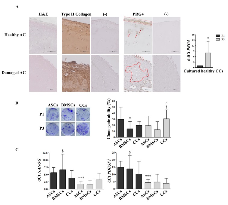
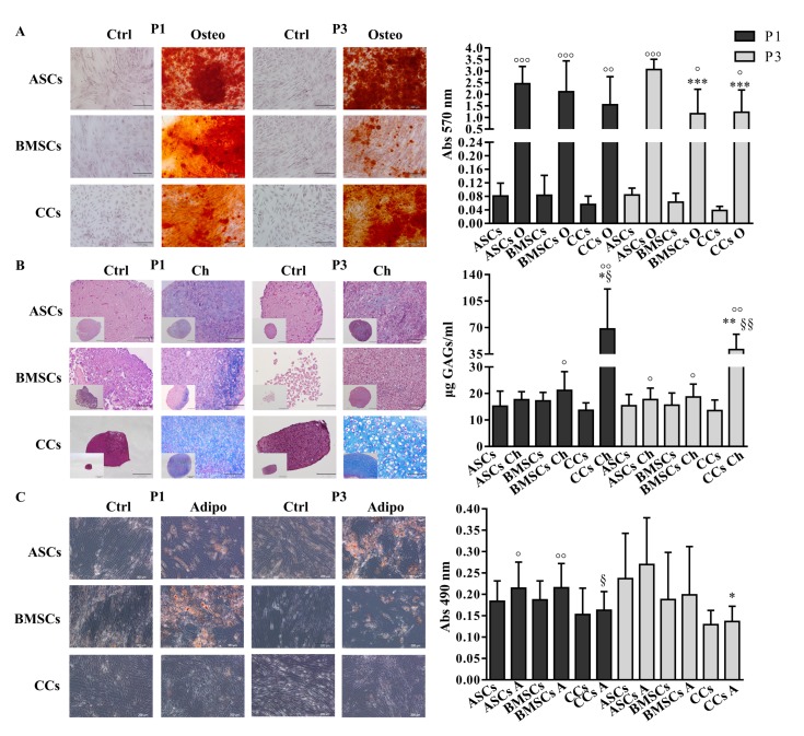
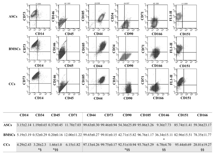
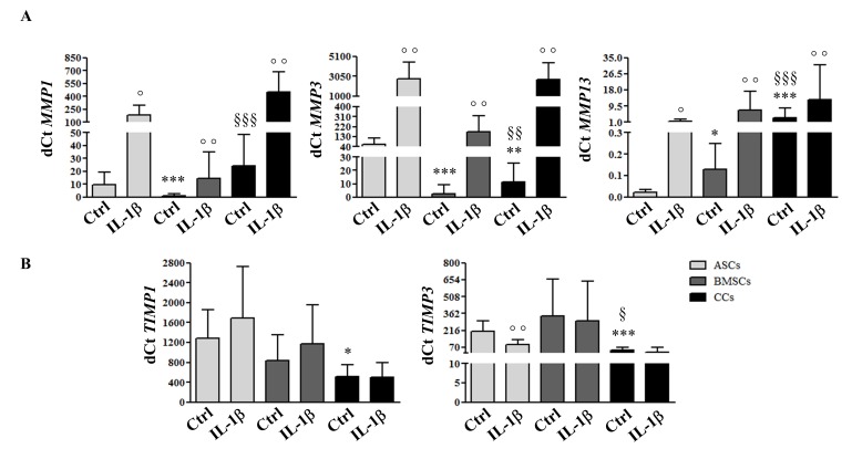
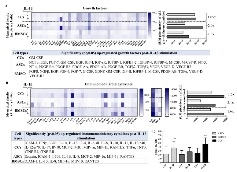
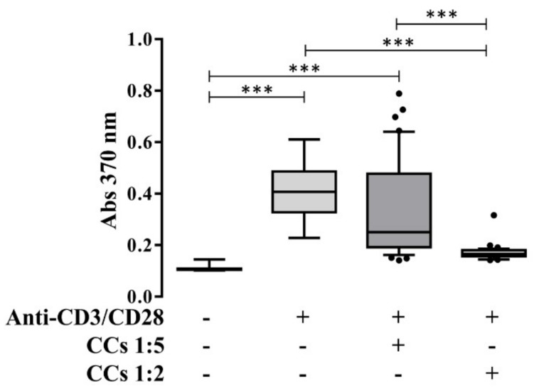
Methods: proteoglycan 4 (PRG4) presence was evaluated in AC and CCs. MSCs and CCs (n = 8) were cultured (P1 to P4) and characterized for clonogenicity, nanog homeobox (NANOG), and POU class 5 homeobox 1 (POU5F1) expression, immunotypification, and tri-lineage differentiation. Their basal and interleukin-1β (IL-1β)-stimulated expression of matrix metalloproteases (MMPs), tissue inhibitors (TIMPs), release of growth factors, and cytokines were analyzed, along with the immunomodulatory ability of CCs.

Results: PRG4 was mainly expressed in the intact AC surface, whereas shifted to the intermediate zone in damaged cartilage and increased its expression in CCs upon culture. All cells exhibited a similar phenotype and stemness maintenance over passages. CCs showed highest chondrogenic ability, no adipogenic potential, a superior basal secretion of growth factors and cytokines, the latter further increased after inflammatory stimulation, and an immunomodulatory behavior. All stimulated cells shared an increased MMP expression without a corresponding TIMP production.

Conclusion: based on the observed features, CCs obtained from pathological joints may constitute a potential tissue-specific therapeutic target or agent to improve damaged cartilage healing, especially damage caused by inflammatory/immune mediated conditions.

Keywords: cartilage cells; cartilage-derived stem/progenitor cells; immunomodulation; inflammation; mesenchymal stem cells; secretome; stemness.


Conflict of interest statement

The authors declare no conflict of interest.

Figures

Figure 1
Figure 1
PRG4 expression, clonogenic ability, and stemness marker expression. (A) Representative immunohistological distribution of type II collagen and PRG4 in healthy and damaged AC (scale bars correspond to 100 µm), and PRG4 expression in culture-expanded CCs (n = 4). (−) indicates negative control (secondary antibody only). (B) Clonogenic ability and (C) stemness marker expression of adipose (ASCs), bone marrow (BMSCs)-derived MSCs and cartilage cells (CCs) obtained from the same eight donors. Cells were analyzed at passage 1 (P1) and passage 3 (P3). * p < 0.05, *** p < 0.001 vs. ASCs at P1, § p < 0.05 vs. BMSCs at P3, ^ p < 0.05 vs. CCs at P1. Data are represented as mean ± SD (n = 8).
Figure 2
Figure 2
Multi-differentiation potential. (A) Osteogenic-O (Alizarin red staining), (B) Chondrogenic-Ch (Alcian blue staining and GAG quantification), and (C) Adipogenic-A (Oil O red staining) differentiation potential assessment. Adipose (ASCs)-, bone marrow (BMSCs)-derived MSCs and cartilage cells (CCs) obtained from the same eight donors were subjected to differentiation at P1 and P3. Scale bars correspond to 200 μm. Only scale bars of the biggest Chondrogenic-Ch images correspond to 100 µm. * p < 0.05, ** p < 0.01, *** p < 0.001 vs. ASCs, § p < 0.05, §§ p < 0.01 vs. BMSCs, ° p < 0.05, °° p < 0.01, °°° p < 0.001 vs. control. Data are represented as mean ± SD (n = 8).
Figure 3
Figure 3
Immunophenotype. Representative expression of the typical pattern of MSC surface markers in all the analyzed cells, with a table reporting the percentages of positive cells for the whole panel of surface markers tested at P4. Adipose (ASCs)-, bone marrow (BMSCs)-derived MSCs, and cartilage cells (CCs). * p < 0.05 vs. ASCs, § p < 0.05, §§ p < 0.05 vs. BMSCs. Data are expressed as mean ± SD (n = 5).
Figure 4
Figure 4
ECM remodeling molecular response to a pro-inflammatory stimulus. Expression of metalloproteases (MMP1, MMP3, MMP13) (A) and their inhibitors (TIMP1, TIMP3) (B) in adipose (ASCs)-, bone marrow (BMSCs)-derived MSCs, and cartilage cells (CCs) obtained from the same donors, assessed by quantitative real-time PCR at P3 before and after IL-1β stimulation. * p < 0.05, ** p < 0.01, *** p < 0.001 vs. ASCs, § p < 0.05, §§ p < 0.01, §§§ p < 0.001 vs. BMSCs, ° p < 0.05, °° p < 0.01 vs. control. Data are expressed as mean ± SD (n = 8).
Figure 5
Figure 5
Secretome multiplex analysis. Secretion of growth factors of (A) inflammation-related cytokines (B) and IL-1Ra (C) in conditioned media obtained from adipose (ASCs)-, bone marrow (BMSCs)-derived MSCs, and cartilage cells (CCs) at basal (−) and post-stimulation with IL-1β (+). Growth factors and cytokines are presented as: overall heat maps of the mean pixel intensity, and arithmetic sum of the pixel intensity calculating the fold increase in the overall secretion from basal to post-stimulation; a table showing significantly upregulated molecules with stimulation (n = 4). For IL-1Ra, °° p < 0.01 vs. control, data are expressed as mean ± SD (n = 8).
Figure 6
Figure 6
PBLs’ proliferation after interaction with CCs. Proliferation of PBLs in presence (+) or absence (−) of anti-CD3/CD28 and IL-1β-treated CCs in a ratio of 1:2 or 1:5 (PBLs:CCs). *** p < 0.001. Data are expressed as mean ± SD (n = 7).

References

    1. Caplan A.I. Adult mesenchymal stem cells: when, where, and how. Stem Cells Int. 2015;2015:628767. doi: 10.1155/2015/628767. - DOI - PMC - PubMed
    1. Namba R.S., Meuli M., Sullivan K.M., Le A.X., Adzick N.S. Spontaneous repair of superficial defects in articular cartilage in a fetal lamb model. J. Bone Joint Surg. Am. 1998;80:4–10. doi: 10.2106/00004623-199801000-00003. - DOI - PubMed
    1. Lietman S.A., Miyamoto S., Brown P.R., Inoue N., Reddi A.H. The temporal sequence of spontaneous repair of osteochondral defects in the knees of rabbits is dependent on the geometry of the defect. J. Bone Joint Surg. Br. 2002;84:600–606. doi: 10.1302/0301-620X.84B4.11631. - DOI - PubMed
    1. Barbero A., Ploegert S., Heberer M., Martin I. Plasticity of clonal populations of dedifferentiated adult human articular chondrocytes. Arthritis Rheum. 2003;48:1315–1325. doi: 10.1002/art.10950. - DOI - PubMed
    1. Dowthwaite G.P., Bishop J.C., Redman S.N., Khan I.M., Rooney P., Evans D.J., Haughton L., Bayram Z., Boyer S., Thomson B., et al. The surface of articular cartilage contains a progenitor cell population. J. Cell Sci. 2004;117:889–897. doi: 10.1242/jcs.00912. - DOI - PubMed