. 2019 Mar 30;18(1):61.
doi: 10.1186/s12943-019-0978-2.

Long noncoding RNA Pvt1 regulates the immunosuppression activity of granulocytic myeloid-derived suppressor cells in tumor-bearing mice

Affiliations

Long noncoding RNA Pvt1 regulates the immunosuppression activity of granulocytic myeloid-derived suppressor cells in tumor-bearing mice

Yu Zheng et al. Mol Cancer. .

Abstract

Background: Myeloid-derived suppressor cells (MDSCs) participate in tumor-elicited immunosuppression by dramatically blocking T-cell-induced antitumor responses, thereby influencing the effectiveness of cancer immunotherapies. Treatments that alter the differentiation and function of MDSCs can partially restore antitumor immune responses. The long noncoding RNA plasmacytoma variant translocation 1 (lncRNA Pvt1) is a potential oncogene in a variety of cancer types. However, whether lncRNA Pvt1 is involved in the regulation of MDSCs has not been thoroughly elucidated to date.

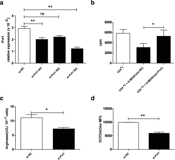
Methods: MDSCs or granulocytic MDSCs (G-MDSCs) were isolated by microbeads and flow cytometry. Bone marrow derived G-MDSCs were induced by IL-6 and GM-CSF. The expression of lncRNA Pvt1 was measured by qRT-PCR. Specific siRNA was used to knockdown the expression of lncRNA Pvt1 in G-MDSCs.

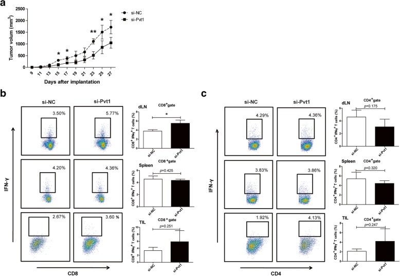
Results: In this study, we found that knockdown of lncRNA Pvt1 significantly inhibited the immunosuppressive function of G-MDSCs in vitro. Additionally, lncRNA Pvt1 knockdown reduced the ability of G-MDSCs to delay tumor progression in tumor-bearing mice in vivo. Notably, lncRNA Pvt1 was upregulated by HIF-1α under hypoxia in G-MDSCs.

Conclusions: Taken together, our results demonstrate a critical role for lncRNA Pvt1 in regulating the immunosuppression activity of G-MDSCs, and lncRNA Pvt1 might thus be a potential antitumor immunotherapy target.

Keywords: Immunosuppression; Long noncoding RNA; Myeloid-derived suppressor cells; Pvt1.


Conflict of interest statement

Ethics approval and consent to participate

This study was approved by the Committee on the Use of Live Animals in Research and Teaching of Jiangsu University.

Consent for publication

Not applicable.

Competing interests

The authors declare that they have no competing interests.

Publisher’s Note

Springer Nature remains neutral with regard to jurisdictional claims in published maps and institutional affiliations.

Figures

Fig. 1
Fig. 1
Pvt1 is highly expressed in tumor-expanded G-MDSCs. A total of 2 × 10^6 Lewis lung carcinoma cells (LLCs) were introduced via s.c. injection into C57BL/6 mice. After 4 weeks, bone marrow cells, splenocytes and a single-cell suspension derived from tumor tissues were collected, and G-MDSCs were later sorted. Splenocytes from wild-type (WT) C57BL/6 mice were collected, and G-MDSCs were isolated. Hierarchical clustering analysis of lncRNAs and protein-coding RNAs that were differentially expressed (fold change > 2) in G-MDSCs sorted from tumor tissue of Lewis tumor-bearing mice and spleens of WT C57BL/6 mice. a Clustering tree for lncRNAs; the expression values are represented in shades of red and green, indicating expression above and below normal values, respectively. b The purity of sorted G-MDSCs was determined via flow cytometry by assessing the expression of two surface markers: Ly6G and CD11b. c The expression level of Pvt1 in total RNA isolated from G-MDSCs from the bone marrow, spleen and tumor tissues of Lewis-bearing mice was measured by qRT-PCR. Fresh G-MDSCs isolated from bone marrow (BM) from WT C57BL/6 mice served as the control. Bone marrow cells (1 × 10^6) from WT C57BL/6 mice were plated in 24-well plates in 1 mL of RPMI 1640 medium containing 10% FBS, 20 ng/mL IL-6 and 20 ng/mL GM-CSF. The cells were then collected, and G-MDSCs were sorted 3 days later. d G-MDSCs cocultured with CFSE-labeled CD4+ T cells at a ratio of 1:1 in the presence of anti-CD3 mAb and anti-CD28 mAb for 72 h. The proliferation of CD4+ T cells was detected by flow cytometry at 488 nm excitation light. e Arg1 activity in G-MDSCs induced from BM cells was measured. f ROS production in G-MDSCs was analyzed via flow cytometry. g The expression level of Pvt1 in G-MDSCs was detected using qRT-PCR. ***p < 0.001, and **p < 0.01; ns: no significance
Fig. 2
Fig. 2
Knockdown of Pvt1 alters the suppressive capacity of G-MDSCs in vitro. G-MDSCs sorted from tumor tissues were obtained from TB mice injected with cells transfected with 50 nM Pvt1 siRNA (si-Pvt1) or negative control (NC) siRNA (si-NC). a qRT-PCR confirmed the efficiency of transfection with si-Pvt1. b G-MDSCs were transfected with Pvt1 siRNA, and then, the cells were harvested after 6 h and cocultured with CD4+T cells at ratio of 1:1 in the presence of anti-CD3 mAb and anti-CD28 mAb for72 h. 3H-thymidine incorporation was used to detect T cells proliferation. c Arg1 activity in G-MDSCs transfected with si-Pvt1 was measured. d ROS production in G-MDSCs was analyzed via flow cytometry. **p < 0.01, and *p < 0.05; ns: no significance; Geo MFI: geometric mean fluorescent intensity
Fig. 3
Fig. 3
c-myc is a potential downstream target of Pvt1 in G-MDSCs. a Scatter plot for protein-coding RNAs. b qRT-PCR was used to detect the mRNA level of c-myc in G-MDSCs sorted from spleen of WT mice and tumor tissues of TB mice. c,d After transfection with si-Pvt1, the mRNA and protein levels of c-myc in G-MDSCs isolated from tumor tissues were measured via qRT-PCR and western blot analyses. **p < 0.01, and *p < 0.05
Fig. 4
Fig. 4
Pvt1 knockdown reduces the ability of G-MDSCs to accelerate tumor progression and inhibit antitumor immune responses. Two groups of mice were given a s.c. injection of a mixture of LLCs and G-MDSCs transfected with si-Pvt1 (si-Pvt1 group) or si-NC (si-NC group). a Tumor volume was measured at the indicated time. b, c The proportions of CD8+IFN-γ+ CTLs and CD4+IFN-γ+ Th1 cells from draining lymph nodes, spleens and tumor tissues were analyzed via flow cytometry. **p < 0.01, and *p < 0.05
Fig. 5
Fig. 5
HIF-1α upregulates Pvt1 expression in G-MDSCs under hypoxic stress. a The mRNA level of HIF-1α in G-MDSCs sorted from spleens and tumor tissues of TB mice was detected using qRT-PCR. G-MDSCs isolated from spleens of TB mice were cultured in an incubator at 37 °C (20% O2, 5% CO2) (normoxic conditions) or in a sealed box containing an anaerobic bag to consume oxygen (O2 < 0.1%, 5% CO2) (hypoxic conditions). b, c The mRNA and protein levels of HIF-1α were measured via qRT-PCR and western blot (WB) analyses, respectively. d Pvt1 expression was analyzed via qRT-PCR. YC-1, a specific inhibitor of HIF-1α, was used to block hypoxia. e, f HIF-1α and (g) Pvt1 expression in the normoxia, hypoxia, and hypoxia+YC-1 groups were detected via qRT-PCR and WB analyses. ***p < 0.001, **p < 0.01, and *p < 0.05; ns: no significance

References

    1. Gabrilovich DI, Nagaraj S. Myeloid-derived suppressor cells as regulators of the immune system[J] Nat Rev Immunol. 2009;9(3):162–174. doi: 10.1038/nri2506. - DOI - PMC - PubMed
    1. Schmielau J, Finn OJ. Activated granulocytes and granulocyte-derived hydrogen peroxide are the underlying mechanism of suppression of t-cell function in advanced cancer patients[J] Cancer Res. 2001;61(12):4756–4760. - PubMed
    1. Tian X, Ma J, Wang T, et al. Long non-coding RNA HOXA transcript antisense RNA myeloid-specific 1-HOXA1 axis downregulates the immunosuppressive activity of myeloid-derived suppressor cells in lung cancer[J] Front Immunol. 2018;9:473. doi: 10.3389/fimmu.2018.00473. - DOI - PMC - PubMed
    1. Veglia F, Perego M, Gabrilovich D. Myeloid-derived suppressor cells coming of age[J] Nat Immunol. 2018;19(2):108–119. doi: 10.1038/s41590-017-0022-x. - DOI - PMC - PubMed
    1. Sinha P, Okoro C, Foell D, et al. Proinflammatory S100 proteins regulate the accumulation of myeloid-derived suppressor cells[J] J Immunol. 2008;181(7):4666–4675. doi: 10.4049/jimmunol.181.7.4666. - DOI - PMC - PubMed

Publication types

MeSH terms

Substances

LinkOut - more resources