Review
. 2019 Mar 30;18(1):59.
doi: 10.1186/s12943-019-0980-8.

Extracellular vesicles in the tumor microenvironment: old stories, but new tales

Affiliations
Review

Extracellular vesicles in the tumor microenvironment: old stories, but new tales

Liu Han et al. Mol Cancer. .

Abstract

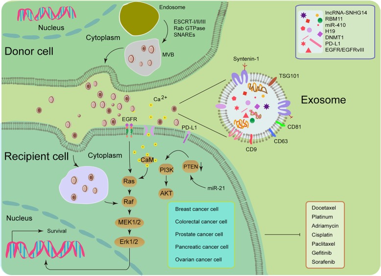
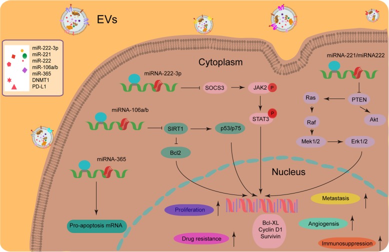
Mammalian cells synthesize and release heterogeneous extracellular vesicles (EVs) which can be generally recognized as subclasses including exosomes, microvesicles (MVs), and apoptotic bodies (ABs), each differing in their biogenesis, composition and biological functions from others. EVs can originate from normal or cancer cells, transfer bioactive cargoes to both adjacent and distant sites, and orchestrate multiple key pathophysiological events such as carcinogenesis and malignant progression. Emerging as key messengers that mediate intercellular communications, EVs are being paid substantial attention in various disciplines including but not limited to cancer biology and immunology. Increasing lines of research advances have revealed the critical role of EVs in the establishment and maintenance of the tumor microenvironment (TME), including sustaining cell proliferation, evading growth suppression, resisting cell death, acquiring genomic instability and reprogramming stromal cell lineages, together contributing to the generation of a functionally remodeled TME. In this article, we present updates on major topics that document how EVs are implicated in proliferative expansion of cancer cells, promotion of drug resistance, reprogramming of metabolic activity, enhancement of metastatic potential, induction of angiogenesis, and escape from immune surveillance. Appropriate and insightful understanding of EVs and their contribution to cancer progression can lead to new avenues in the prevention, diagnosis and treatment of human malignancies in future medicine.

Keywords: Cancer biology; Clinical biomarker; Extracellular vesicles; Therapeutic target; Tumor microenvironment.


Conflict of interest statement

Ethics approval and consent to participate

Not applicable.

Consent for publication

Not applicable.

Competing interests

The authors declare that they have no competing interests.

Publisher’s Note

Springer Nature remains neutral with regard to jurisdictional claims in published maps and institutional affiliations.

Figures

Fig. 1
Fig. 1
Illustrative diagram for exosome-mediated transfer of therapeutic resistance in the tumor microenvironment (TME). Drug-resistant (donor) cells may communicate with drug-sensitive (recipient) cells by the intercellular transfer of various types of EVs, such as exosomes (usually expressing tetraspanins such as CD9/63/81, TSG101 and syntenin-1), which are of endocytic origin [124]. Upon fusion of secretory multivesicular bodies (MVBs) with the plasma membrane, exosomes are released into the extracellular space. The initial steps of this process are usually modulated by the endosomal sorting complex required for transport (ESCRT) [125]. The mechanisms involved in the release of exosomes are also regulated by other protein families, such as Rab GTPases and SNARES [125, 126]. Once EVs reach the recipient cells, they may fuse with their plasma membrane or be internalized by the endocytic pathway. Exosomes may transfer miRNAs, lncRNAs, proteins (such as drug-efflux pumps), and other key players responsible for drug resistance, which allows de novo development or horizontal dissemination of cancer resistance traits to the recipient cell populations. For example, mesenchymal stem cell (MSC)-derived exosomes trigger the activation of calcium-dependent protein kinases and EGFR/Ras/Raf/Mek/Erk kinase cascade in gastric cancer cells, while polarized macrophages promote cisplatin resistance of gastric cancer cells by exosomal transfer of miR-21 which functionally activates PI3K/AKT signaling via down-regulation of PTEN in recipient cells [127, 128]
Fig. 2
Fig. 2
Multiple roles of EV-delivered cargoes such as microRNAs (miRNAs) in altering the phenotypes of recipient cancer cells and shaping a pathologically active tumor microenvironment (TME). Cancer cells and stromal cells utilize EVs such as exosomes to influence surrounding cells within the microenvironmental niche by transferring bioactive molecules including miRNAs. Sorting miRNAs to EVs is regulated by cell activation-dependent changes in miRNA levels within donor cells. Specifically, miRNA-365, miRNA-106a/b, miRNA-222-3p and miRNA-221/222 are not only overexpressed in donor cells but also enriched in their exosomes, and upon exosome-mediated transmission these miRNAs can significantly enhance resistance of recipient cancer cells against anticancer agents [–133]. In addition, other malignant properties including but not limited to proliferation capacity, angiogenesis capability, metastatic potential and immunosurveillance evasion are also subject to the impact of EVs released by stromal or cancer cells in the TME

References

    1. Ellis TN, Kuehn MJ. Virulence and immunomodulatory roles of bacterial outer membrane vesicles. Microbiol Mol Biol Rev. 2010;74:81–94. doi: 10.1128/MMBR.00031-09. - DOI - PMC - PubMed
    1. Beveridge TJ. Structures of gram-negative cell walls and their derived membrane vesicles. J Bacteriol. 1999;181:4725–4733. - PMC - PubMed
    1. Esselman WJ, Miller HC. Modulation of B cell responses by glycolipid released from antigen-stimulated T cells. J Immunol. 1977;119:1994–2000. - PubMed
    1. Freimuth WW, Miller HC, Esselman WJ. Soluble factors containing Thy-1 antigen shed from lymphoblastoid cells modulate in vitro plaque-forming cell response. J Immunol. 1979;123:201–208. - PubMed
    1. Harding C, Heuser J, Stahl P. Receptor-mediated endocytosis of transferrin and recycling of the transferrin receptor in rat reticulocytes. J Cell Biol. 1983;97:329–339. doi: 10.1083/jcb.97.2.329. - DOI - PMC - PubMed

Publication types