Review
. 2019 Mar 30;18(1):62.
doi: 10.1186/s12943-019-0967-5.

The roles of extracellular vesicles in gastric cancer development, microenvironment, anti-cancer drug resistance, and therapy

Affiliations
Review

The roles of extracellular vesicles in gastric cancer development, microenvironment, anti-cancer drug resistance, and therapy

Tingting Huang et al. Mol Cancer. .

Abstract

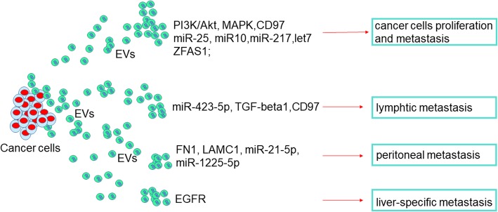
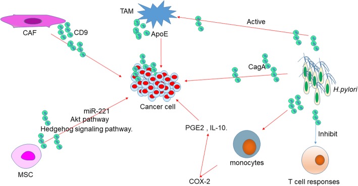
Gastric cancer (GC) is one of the leading causes of cancer-related death in both men and women due to delayed diagnosis and high metastatic frequency. Extracellular vesicles (EVs) are membrane-bound nanovesicles which are released by cells into body fluids such as plasma, saliva, breast milk, cerebrospinal fluid, semen, urine, lymphatic fluid, amniotic fluid, sputum and synovial fluid. EVs deliver almost all types of biomolecules such as proteins, nucleic acids, metabolites, and even pharmacological compounds. These bioactive molecules can be delivered to recipient cells to influence their biological properties, modify surrounding microenvironment and distant targets. The extensive exploration of EVs enhances our comprehension of GC biology referring to tumor growth, metastasis, immune response and evasion, chemoresistance and treatment. In this review, we will sum up the effects of GC-derived EVs to the tumor microenvironment. Moreover, we will also summarize the function of microenvironment-derived EVs in GC and discuss how the bidirectional communication between tumor and microenvironment affect GC growth, metastatic behavior, immune response, and drug resistance. At last, we prospect the clinical application viewpoint of EVs in GC.

Keywords: Drug resistance; Exosomes; Extracellular vesicles; Gastric cancer; Tumor microenvironment.


Conflict of interest statement

Ethics approval and consent to participate

Not applicable.

Consent for publication

Yes.

Competing interests

The authors declare that they have no competing interests.

Publisher’s Note

Springer Nature remains neutral with regard to jurisdictional claims in published maps and institutional affiliations.

Figures

Fig. 1
Fig. 1
Release of EVs and its contents. Primarily, the EVs are originally derived from lysosomes and late endosomes. Then, they can be released into the extracellular environment. The contents of EVs, which contain DNAs, mRNAs, small RNAs, and proteins can be transferred from the original cell to their target cells in local microenvironment or at distant site that can possibly give rise to intercellular communication networks. Abbreviations: EVs, extracellular vesicles
Fig. 2
Fig. 2
Functions of cancer derived EVs in GC progression and metastasis. The first general mechanism is that GC cells-derived EVs promote tumor cells growth and metastasis through overexpression of multiple proteins, miRNAs and LncRNAs. The second general mechanism is that metastasis, including lymphtic, peritoneal, and liver-specific metastasis, which can be induced by tumor-derived EVs via different pathways in GC. Abbreviations: EGFR, epidermal growth factor receptor
Fig. 3
Fig. 3
The functional network of cancer derived EVs in GC microenvironment. GC cells-derived EVs promote angiogenesis via releasing miR-130a. Pericytes, MSCs, and fibroblasts absorbed EVs to induce CAFs transformation in tumor microenvironment through different pathway or miRNAs in cells. The functions of cancer cells-derived EVs in adipocytes differentiation. Different immune cells in tumor microenvironment can be affected by tumor-derived EVs. GC-derived EVs inhibit T cell immunity, polarize neutrophils to a pro-tumor phenotype, induce macrophages to release more proinflammatory factors and active Th17 to promote cancer progression. Abbreviations: GC, gastric cancer; MSC, mesenchymal stem cell; CAF, cancer-associated fibroblast
Fig. 4
Fig. 4
The regulation network of microenvironment-derived EVs as well as H.pylori-derived EVs in GC. EVs secreted by CAF, MSC, and TAM induce GC progression through different pathways and molecules. H.pylori releases CagA-containing EVs and other EVs that inhibit T cell responses, active monocytes to induce COX-2 expression, and active TAM to induce gastric carcinogenesis. Abbreviations: TAM, tumor-associated macrophage; CAF, cancer-associated fibroblast; MSC, mesenchymal stem cell

References

    1. Ferlay J, Soerjomataram I, Dikshit R, Eser S, Mathers C, Rebelo M, Parkin DM, Forman D, Bray F. Cancer incidence and mortality worldwide: sources, methods and major patterns in GLOBOCAN 2012. Int J Cancer. 2015;136. - PubMed
    1. Cancer Genome Atlas Research N Comprehensive molecular characterization of gastric adenocarcinoma. Nature. 2014;513:202. doi: 10.1038/nature13480. - DOI - PMC - PubMed
    1. Schorey JS, Bhatnagar S. Exosome function: from tumor immunology to pathogen biology. Traffic (Copenhagen, Denmark) 2008;9:871–881. doi: 10.1111/j.1600-0854.2008.00734.x. - DOI - PMC - PubMed
    1. Mathivanan S, Ji H, Simpson RJ. Exosomes: Extracellular organelles important in intercellular communication. J Proteome. 2010;73:1907–1920. doi: 10.1016/j.jprot.2010.06.006. - DOI - PubMed
    1. Skog J, Würdinger T, van Rijn S, Meijer DH, Gainche L, Sena-Esteves M, Curry WT, Jr, Carter BS, Krichevsky AM, Breakefield XO. Glioblastoma microvesicles transport RNA and proteins that promote tumour growth and provide diagnostic biomarkers. Nat Cell Biol. 2008;10:1470–1476. doi: 10.1038/ncb1800. - DOI - PMC - PubMed

Publication types

MeSH terms

Substances