. 2019 Apr 16;116(16):8000-8009.
doi: 10.1073/pnas.1817183116. Epub 2019 Mar 29.

Loss of postnatal quiescence of neural stem cells through mTOR activation upon genetic removal of cysteine string protein-α

Affiliations

Loss of postnatal quiescence of neural stem cells through mTOR activation upon genetic removal of cysteine string protein-α

Jose L Nieto-González et al. Proc Natl Acad Sci U S A. .

Abstract

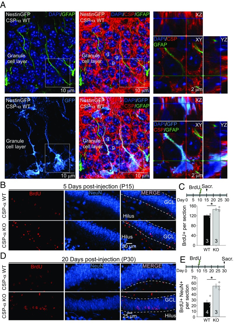
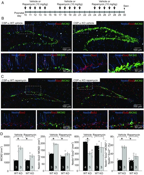
Neural stem cells continuously generate newborn neurons that integrate into and modify neural circuitry in the adult hippocampus. The molecular mechanisms that regulate or perturb neural stem cell proliferation and differentiation, however, remain poorly understood. Here, we have found that mouse hippocampal radial glia-like (RGL) neural stem cells express the synaptic cochaperone cysteine string protein-α (CSP-α). Remarkably, in CSP-α knockout mice, RGL stem cells lose quiescence postnatally and enter into a high-proliferation regime that increases the production of neural intermediate progenitor cells, thereby exhausting the hippocampal neural stem cell pool. In cell culture, stem cells in hippocampal neurospheres display alterations in proliferation for which hyperactivation of the mechanistic target of rapamycin (mTOR) signaling pathway is the primary cause of neurogenesis deregulation in the absence of CSP-α. In addition, RGL cells lose quiescence upon specific conditional targeting of CSP-α in adult neural stem cells. Our findings demonstrate an unanticipated cell-autonomic and circuit-independent disruption of postnatal neurogenesis in the absence of CSP-α and highlight a direct or indirect CSP-α/mTOR signaling interaction that may underlie molecular mechanisms of brain dysfunction and neurodegeneration.

Keywords: DNAJC5; adult neurogenesis; adult-onset neuronal ceroid lipofuscinosis; lysosome; synaptic neurodegeneration.


Conflict of interest statement

The authors declare no conflict of interest.

Figures

Fig. 1.
Fig. 1.
Postnatal increase of newborn neurons at the hippocampal neurogenic niche in CSP-α KO mice. (A, Left and Center) Maximum intensity projections of a z-stack of confocal images from nestin-GFP transgenic CSP-α WT mouse hippocampal slices labeled with DAPI (dark blue), anti-GFP (light blue), and anti-GFAP (green) antibodies. (A, Right) Areas within dashed-line squares are magnified, showing three optical sections [XY (Middle), XZ (Upper), and YZ (Right)] of RGLs in the dentate gyrus identified by the colocalization of nestin (blue) and GFAP (green). Better visualization is provided in Movie S1. We have found that 100% of nestin+/GFAP+ cells express CSP-α (28 cells observed in three different mice). (B) Maximum intensity projection of a z-stack of confocal images of CSP-α WT and KO hippocampal slices labeled with antibodies against BrdU (red) and NeuN (blue) 5 d after BrdU injection. (C) Increased number of BrdU+ nuclei per section in CSP-α KO mice compared with WT mice 5 d postinjection (four sections per mouse for WT and four to five sections per mouse for CSP-α KO; n = 3 for each genotype). Sacr., sacrifice. (D) Maximum intensity projection of a z-stack of confocal images of CSP-α WT and KO hippocampal slices labeled with antibodies against BrdU (red) and NeuN (blue) 20 d after BrdU injection. (E) Increased number of newborn neurons identified as BrdU+ nuclei per section colocalizing with NeuN in CSP-α KO mice at 20 d postinjection [four sections per mouse for WT (n = 4) and four and five sections per mouse for CSP-α KO (n = 3)]. Numbers in bars indicate the number of mice used. Mean ± SEM (*P < 0.05, Student’s t test).
Fig. 2.
Fig. 2.
Quiescence loss and RGL pool depletion in the dentate gyrus of CSP-α KO mice. (A) Maximum intensity projection of a z-stack of confocal images of CSP-α WT and KO hippocampal dentate gyri labeled with antibodies against nestin (blue), Sox2 (red), and MCM2 (green) at P15. (B) Magnification of the area enclosed within the dashed-line squares in A. (C) At P15, no significant differences are apparent in the size of the RGL cell pool (nestin+, Sox+ cells per square millimeter), but the number (nestin+, Sox+, MCM2+ cells per square millimeter) and percentage of dividing RGL cells are strongly increased in CSP-α KO mice compared with WT controls (five to six sections per mouse for WT and five to six sections per mouse for CSP-α KO; n = 3 for each genotype). (D) Maximum intensity projection of a z-stack of confocal images of CSP-α WT and KO hippocampal dentate gyri labeled with antibodies against nestin (blue), Sox2 (red), and MCM2 (green) at P30. (E) Magnification of the area enclosed within the dashed-line squares in D. (F) At P30, the RGL cell pool size (nestin+, Sox+ cells per square millimeter) is clearly reduced and the number (nestin+, Sox+, MCM2+ cells per square millimeter) and percentage of dividing RGL cells are strongly increased in CSP-α KO mice compared with WT controls (six to seven sections per mouse for WT and seven sections per mouse for CSP-α KO; n = 3 for each genotype). Numbers in bars indicate the number of mice used. Mean ± SEM (*P < 0.05, Student’s t test).
Fig. 3.
Fig. 3.
Hyperactivation of the mTORC1 signaling pathway causes hyperproliferation in CSP-α KO neurospheres. (A) Rapamycin (25 nM) strongly inhibited the phosphorylation of ribosomal protein S6 in WT and CSP-α KO neurosphere cultures (n = 3 cultures from three mice for each genotype). (B) Typical images of WT and CSP-α KO cultures incubated in vehicle or rapamycin (25 nM) for 7 d starting from the time at which cells were cultured. (C) Incubation in the presence of rapamycin (25 nM), compared with incubation in vehicle, reestablished the normal neurosphere size in CSP-α KO cultures (mean value of P < 0.05, Student’s t test). Rapamycin decreased the size of both WT and CSP-α KO neurospheres.
Fig. 4.
Fig. 4.
Rescue of neural stem cell quiescence in vivo upon pharmacological inhibition of the mTORC1-dependent signaling pathway in CSP-α KO mice. (A) Time line for treatment with vehicle or rapamycin (10 mg/kg). Sacr., sacrifice. (B) Maximum intensity projection of a z-stack of confocal images of CSP-α WT and KO hippocampal dentate gyri labeled with antibodies against nestin (blue), Sox2 (red), and MCM2 (green) from 30-d-old mice treated with vehicle. Magnified views (Lower) of the areas in dashed squares (Upper) show the notable increase in neurogenic proliferation (MCM2+ cells in green, arrowheads) in CSP-α KO mice. (C) Confocal images, as in A, taken from 30-d-old mice treated with rapamycin (10 mg/kg, administered once per day for 5 consecutive days per week for ∼20 d). Magnified views (Lower) of areas in dashed squares (Upper) show the strong reduction in the number of MCM2+ cells (green, arrowheads) in CSP-α KO mice, indicating the normalization of stem cell proliferation upon pharmacological inhibition of the mTORC1-dependent signaling pathway. (D) Rapamycin reestablishes the total number of proliferating cells (MCM2+ cells; Left), the number of dividing RGL stem cells (nestin+, Sox2+, MCM2+ cells; Center), and the percentage of activated proliferating RGL stem cells (Right) (four to six sections per mouse for WT and KO; n = 3 for each genotype, except for quantification of nestin+, Sox2+ cells of mice treated with vehicle) to WT levels. Numbers in bars indicate the number of mice used. Values represent mean ± SEM (*P < 0.05, two-way ANOVA).
Fig. 5.
Fig. 5.
Increased proliferation and loss of quiescence in RGLs lacking CSP-α in the dentate gyrus of the Sox2CreERT2:Dnajc5flox/− conditional mouse model. (A) Genomic strategy for cre-recombinase-dependent deletion at Dnajc5 mouse locus. (B) Timeline for treatment with tamoxifen (TMX; 75 mg/kg) and EdU (50 mg/kg). (C) Maximum intensity projection of a z-stack of confocal images of slices labeled with EdU and TO-PRO-3. Slices were obtained from Sox2CreERT2:Dnajc5flox/+ animals injected with vehicle and Sox2CreERT2:Dnajc5flox/− animals injected with TMX (75 mg/kg). (D) Increased number of EdU-positive nuclei per section in Sox2CreERT2:Dnajc5flox/+:vehicle compared with Sox2CreERT2:Dnajc5flox/−:TMX animals at 2 d postinjection (dpi) of EdU (6–7 sections/mouse, n = 3 for each genotype). (E) Maximum intensity projections of a z-stack of confocal images of Sox2CreERT2:Dnajc5flox/+:vehicle and Sox2CreERT2:Dnajc5flox/−:TMX hippocampal dentate gyri labeled with antibodies against nestin (blue), Sox2 (red), and MCM2 (green) are shown at P42. (F) Magnification of the area enclosed within the dashed-line square in C. (G) Size of the RGL cell pool (nestin+, Sox2+ cells per square millimeter; six to seven sections per mouse; n = 4 for each genotype) is significantly decreased, and the percentage of dividing RGL cells (six to seven sections per mouse; n = 3 for each genotype) is significantly increased in Sox2CreERT2:Dnajc5flox/−:vehicle mice compared with Sox2CreERT2:Dnajc5flox/:TMX mice. No differences were found in the number of dividing RGL cells (nestin+, Sox2+, MCM2+ cells per square millimeter; six to seven sections per mouse; n = 3 for each genotype). Quantifications after tamoxifen treatment for 12 d. *P < 0.05. White circles correspond to values from individual mice.

References

    1. Altman J, Das GD. Autoradiographic and histological evidence of postnatal hippocampal neurogenesis in rats. J Comp Neurol. 1965;124:319–335. - PubMed
    1. Nakashiba T, et al. Young dentate granule cells mediate pattern separation, whereas old granule cells facilitate pattern completion. Cell. 2012;149:188–201. - PMC - PubMed
    1. Clelland CD, et al. A functional role for adult hippocampal neurogenesis in spatial pattern separation. Science. 2009;325:210–213. - PMC - PubMed
    1. Akers KG, et al. Hippocampal neurogenesis regulates forgetting during adulthood and infancy. Science. 2014;344:598–602. - PubMed
    1. Christian KM, Song H, Ming GL. Functions and dysfunctions of adult hippocampal neurogenesis. Annu Rev Neurosci. 2014;37:243–262. - PMC - PubMed

Publication types