. 2019 Apr 16;116(16):7926-7931.
doi: 10.1073/pnas.1820892116. Epub 2019 Mar 29.

Small-molecule factor B inhibitor for the treatment of complement-mediated diseases

Affiliations

Small-molecule factor B inhibitor for the treatment of complement-mediated diseases

Anna Schubart et al. Proc Natl Acad Sci U S A. .

Abstract

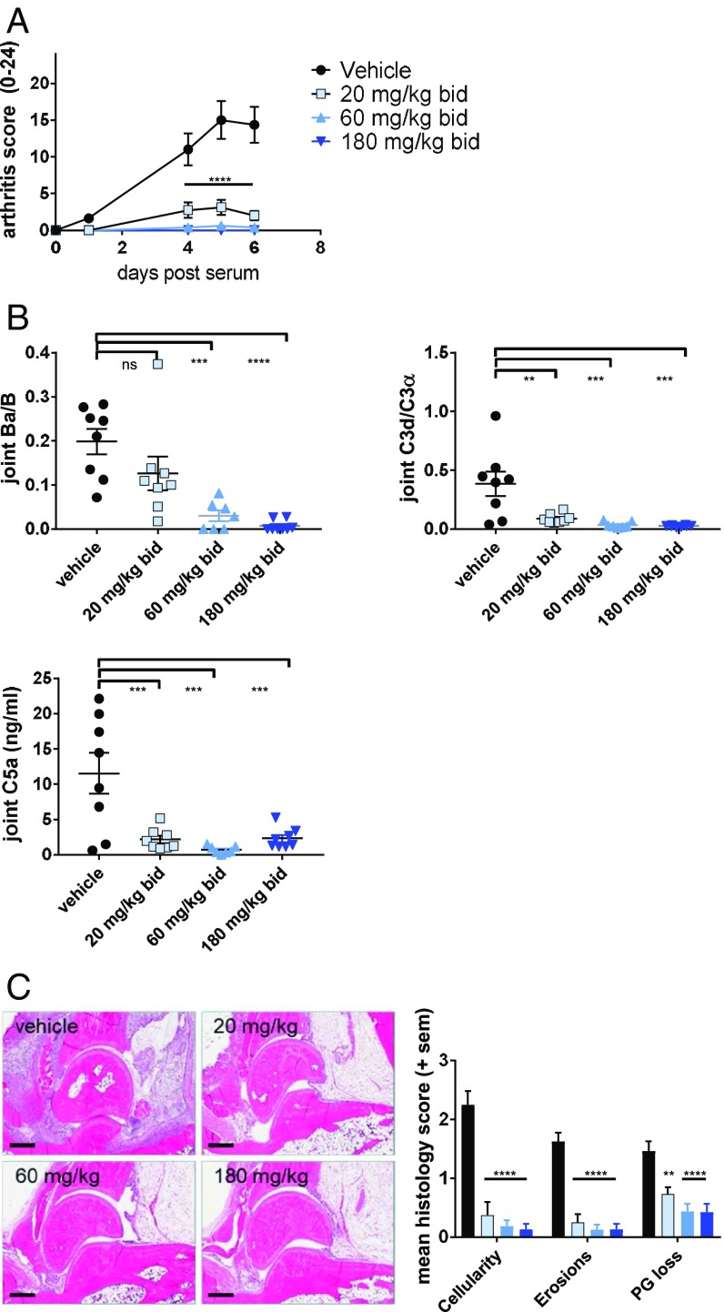
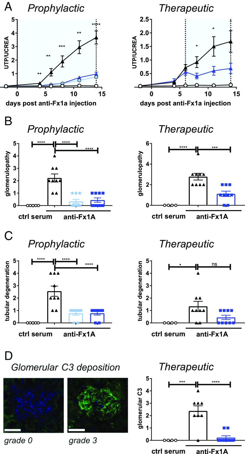
Dysregulation of the alternative complement pathway (AP) predisposes individuals to a number of diseases including paroxysmal nocturnal hemoglobinuria, atypical hemolytic uremic syndrome, and C3 glomerulopathy. Moreover, glomerular Ig deposits can lead to complement-driven nephropathies. Here we describe the discovery of a highly potent, reversible, and selective small-molecule inhibitor of factor B, a serine protease that drives the central amplification loop of the AP. Oral administration of the inhibitor prevents KRN-induced arthritis in mice and is effective upon prophylactic and therapeutic dosing in an experimental model of membranous nephropathy in rats. In addition, inhibition of factor B prevents complement activation in sera from C3 glomerulopathy patients and the hemolysis of human PNH erythrocytes. These data demonstrate the potential therapeutic value of using a factor B inhibitor for systemic treatment of complement-mediated diseases and provide a basis for its clinical development.

Keywords: alternative pathway; complement; drug discovery; factor B; nephropathy.


Conflict of interest statement

Conflict of interest statement: Novartis Pharma AG has filed patent applications on the compounds. A.S., K.A., N.M., H.S., T.E., C.M.A., A.M.S., S.-M.L., M.C., A.L.-E., S.S., G.W., L.P., V.D., T.F., R.G.K., C.Z., E.V., F.S., B.G., P.E., N.H., T.M.S., F.C., U.A.A., B.H., M.M., R.S., C.W., B.J., J.M., S.F., R.H., and J.E. are or were employees of Novartis Pharma AG during this work.

Figures

Fig. 1.
Fig. 1.
Identification and structure-based optimization of a potent and selective FB inhibitor. (A) Chemical structure of compound 1, compound 2, and LNP023 and corresponding IC50 values for inhibition of FB. (B) Cocrystal structure of human FB in complex with compound 1 (yellow sticks) at 1.64 Å resolution. (C) Overlay of compound 1 (yellow sticks) and compound 2 (white sticks) with close-up view into the S1 pocket. (D) Overlay of compound 2 (white sticks) and LNP023 (cyan sticks). A sulfate ion (yellow) from the cocrystal structure of compound 2 is shown. The catalytic residues His57 and Ser195 and other key residues of the ligand binding pockets are highlighted and shown as sticks.
Fig. 2.
Fig. 2.
LNP023 blocks KRN-induced arthritis. Arthritis was induced by KRN/I-Ag7 serum transfer in C57BL/6 mice 1 h after the first dose of compound (n = 8 mice per dose group). (A) Disease score was measured by joint swelling and shown as mean ± SEM. (B) Levels of complement activation products Ba, C3d, and C5a in the joint. (C) Representative images of joint histological analysis (H&E stain) and evaluation of inflammatory cell infiltration, bone erosion, and proteoglycan (PG) loss (safranin O staining) after 6 d of treatment. Vehicle (black bars), 20 mg/kg b.i.d. (light blue bars), 60 mg/kg b.i.d. (medium blue bars), and 180 mg/kg b.i.d. (dark blue bars). Data are mean ± SEM. P values were calculated using one-way ANOVA followed by Dunnett’s multiple comparison test with a single pooled variance (for complement activation fragments and histology) or repeat measure two-way ANOVA followed by Dunnett’s multiple comparison test; **P < 0.01, ***P < 0.001, ****P < 0.0001. (Scale bars, 400 µm.)
Fig. 3.
Fig. 3.
Efficacy of LNP023 in an experimental model of membranous nephropathy. Passive Heymann nephritis was induced by injection of an anti-fraction 1A (anti-Fx1A) antibody serum, and LNP023 was dosed either prophylactically starting at day 0 or therapeutically starting at day 6 after disease onset. Nine animals were used per treatment group, and five animals received control (ctrl) serum. Light blue bars/symbols, 20 mg/kg b.i.d.; dark blue bars/symbols, 60 mg/kg b.i.d. (A) Proteinuria as determined by the urinary ratio of total protein (UTP) and creatinine (UCREA). The light blue areas indicate the duration of compound dosing. Open circles, control serum; black triangles, vehicle; blue filled circles, LNP023 20 mg/kg; blue triangles, LNP023 60 mg/kg. (B) Glomerulopathy (as characterized by enlarged glomeruli, basal membrane and Bowman’s capsule thickening, and enlarged/rounded podocytes) and (C) tubular degeneration were determined by histopathological analysis after H&E staining on day 14 (therapeutic dosing) or day 15 (prophylactic dosing). (D) Glomerular C3 deposition. (Left) Representative histological staining of glomerular C3 deposition (grade 0 and 3). (Scale bars, 50 µm.) (Right) Glomerular C3 deposition after therapeutic dosing of LNP023 (day 15). Data are mean ± SEM. P values were calculated using one-way ANOVA followed by Dunnett’s multiple comparison test with a single pooled variance (for complement activation fragments and histology) or repeat measure two-way ANOVA followed by Dunnett’ multiple comparison test; *P < 0.05, **P < 0.01, ***P < 0.001, ****P < 0.0001.
Fig. 4.
Fig. 4.
LNP023 blocks complement activity in the serum of C3G patients and prevents lysis of human PNH erythrocytes. (A) Inhibition of C3 convertase activity in serum of C3G patients. Patient serum was incubated with normal human serum, and C3 degradation was detected by immunofixation electrophoresis and quantified. The AP is blocked in the presence of EDTA (maximum inhibition control) but not in the presence of Mg2+-EGTA, which blocks the classical and lectin pathways. LNP023 is shown in light blue at 1.1 µM and dark blue at 3.3 µM, and an FD inhibitor is shown in green. (B) Prevention of C3G patient serum-induced hemolysis of sheep erythrocytes. Erythrocytes were incubated with C3G patient serum and LNP023 (0.15 µM) or an FD inhibitor (0.15 µM). Hemolysis was measured by increase in optical density at 415 nm. Addition of EDTA and EGTA was used as negative and positive control, respectively. (C) Inhibition of nephritic factor-stabilized C3 convertase activity. Sheep erythrocytes coated with preformed C3 convertase were incubated with total IgGs from C3G patient sera in the absence or presence of LNP023 (0.15 µM) or an FD inhibitor (0.15 µM). C3 convertase stabilized at 15 min was calculated as % Z (Z15min/Z0min × 100). The assay was adjusted to yield one active C3 convertase per sheep erythrocyte (Z0min = 1; SI Appendix). (D) Inhibition of hemolysis of PNH erythrocytes. Blood from three patients was analyzed in 2–14 repeats, mean ± SEM. Inhibitors were used at 1 µM. Hemolysis of PNH erythrocytes was determined by FACS analysis. (E) Dose–response curves for inhibition of hemolysis of erythrocytes from PNH patients for LNP023, FD inhibitor, and anti-C5 antibody. Hemolysis was measured by FACS analysis. Curves are representatives of three patients measured in 2–14 repeats. For each curve, the average per person from all repeats was used. P values were calculated using one-way ANOVA followed by Dunnett’s multiple comparison test with a single pooled variance; *P < 0.05, **P < 0.01, ***P < 0.001, ****P < 0.0001.

References

    1. Walport MJ. Complement. First of two parts. N Engl J Med. 2001;344:1058–1066. - PubMed
    1. Ricklin D, Hajishengallis G, Yang K, Lambris JD. Complement: A key system for immune surveillance and homeostasis. Nat Immunol. 2010;11:785–797. - PMC - PubMed
    1. Schramm EC, et al. Genetic variants in the complement system predisposing to age-related macular degeneration: A review. Mol Immunol. 2014;61:118–125. - PMC - PubMed
    1. Brodsky RA. Paroxysmal nocturnal hemoglobinuria. Blood. 2014;124:2804–2811. - PMC - PubMed
    1. Rodríguez de Córdoba S, Hidalgo MS, Pinto S, Tortajada A. Genetics of atypical hemolytic uremic syndrome (aHUS) Semin Thromb Hemost. 2014;40:422–430. - PubMed