. 2019 Mar 29;10(1):1424.
doi: 10.1038/s41467-019-09416-2.

Specialized dendritic cells induce tumor-promoting IL-10+IL-17+ FoxP3neg regulatory CD4+ T cells in pancreatic carcinoma

Affiliations

Specialized dendritic cells induce tumor-promoting IL-10+IL-17+ FoxP3neg regulatory CD4+ T cells in pancreatic carcinoma

Rocky M Barilla et al. Nat Commun. .

Abstract

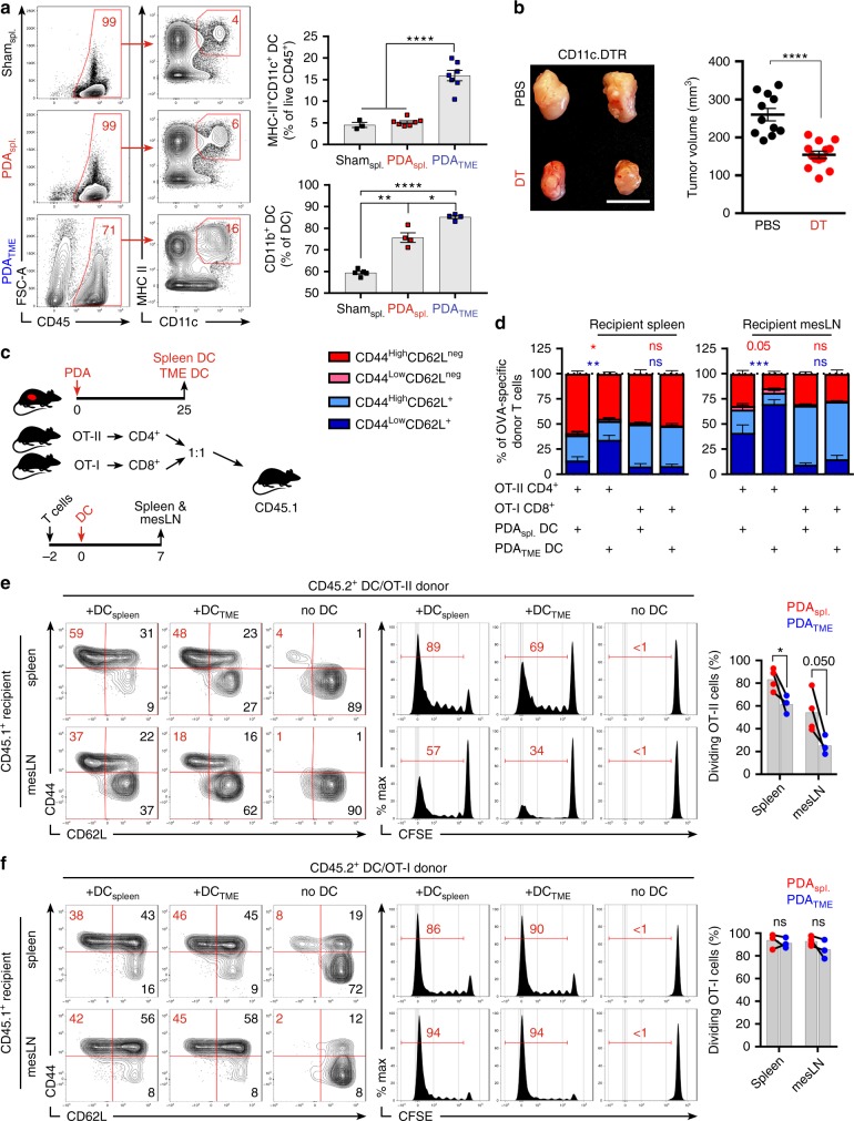
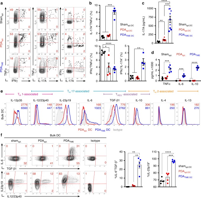
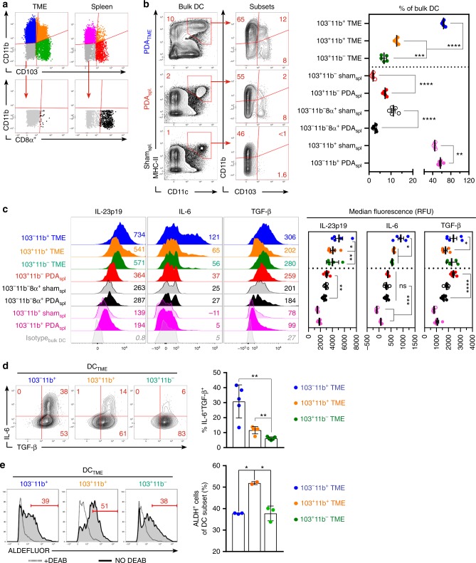
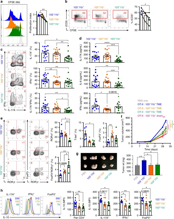
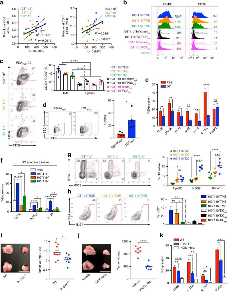
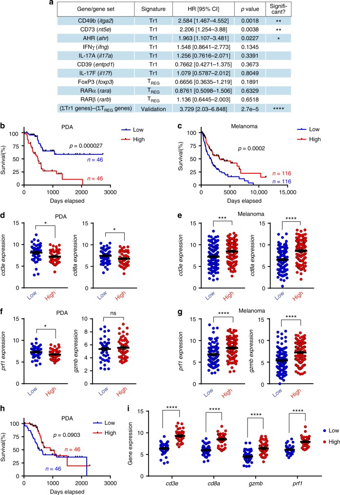
The drivers and the specification of CD4+ T cell differentiation in the tumor microenvironment and their contributions to tumor immunity or tolerance are incompletely understood. Using models of pancreatic ductal adenocarcinoma (PDA), we show that a distinct subset of tumor-infiltrating dendritic cells (DC) promotes PDA growth by directing a unique TH-program. Specifically, CD11b+CD103- DC predominate in PDA, express high IL-23 and TGF-β, and induce FoxP3neg tumor-promoting IL-10+IL-17+IFNγ+ regulatory CD4+ T cells. The balance between this distinctive TH program and canonical FoxP3+ TREGS is unaffected by pattern recognition receptor ligation and is modulated by DC expression of retinoic acid. This TH-signature is mimicked in human PDA where it is associated with immune-tolerance and diminished patient survival. Our data suggest that CD11b+CD103- DC promote CD4+ T cell tolerance in PDA which may underscore its resistance to immunotherapy.


Conflict of interest statement

G.M. has research agreements with GSK, Pfizer, and Puretech Health. The remaining authors declare no competing interests.

Figures

Fig. 1
Fig. 1
Distinct pancreatic ductal adenocarcinoma (PDA)-infiltrating dendritic cell (DC) promote tumor progression and regulate CD4+ T-cell differentiation. a CD45+ cells from the spleen or tumor of orthotopic PDA-bearing mice or from the spleen of control mice were gated using flow cytometry and tested for co-expression of CD11c and MHCII. CD11c+MHCII+ cells were then sub-gated and tested for expression of CD11b. Representative contour plots and quantitative data are shown. This experiment was repeated >5 times. b WT mice were made chimeric using bone marrow from CD11c.DTR mice. Cohorts were challenged with orthotopic PDA 7 weeks later. On day 12, mice began serial treatment with diphtheria toxin (DT) or phosphate-buffered saline (PBS) before sacrifice on day 25. Representative gross images of tumors and quantitative analysis of tumor volume are shown. This experiment had similar results for >5 times (scale bars = 1 µm). cf PDATME DC were harvested on day 25 from tumor-bearing mice, loaded with either Ova323–339 or Ova257–264 peptide and administered in equal number i.p. to CD45.1 mice that had been transferred i.v. 2 days prior with equal numbers of CFSE-labeled OT-I and OT-II T cells. Parallel experiments were performed using PDAspl. DC. On day 7 after either PDATME or PDAspl DC.Ova administration, spleen and mesenteric lymph nodes (LNs) were harvested and CD45.2+ CD4+ and CD8+ T cells tested for co-expression of CD44 and CD62L and dilution of CFSE. c A schematic of the experimental regimen is depicted. d Quantitative results and e representative flow cytometry data for CD4+ T cells and f CD8+ T cells are shown. This experiment was repeated twice (*p < 0.05, **p < 0.01, ***p < 0.001, ****p < 0.0001, t-test), SEM
Fig. 2
Fig. 2
PDATME dendritic cell (DC) exhibit a unique cytokine profile and promote IL-17 expression in naive CD4+ T cells. ac Ova323–339 peptide-pulsed PDATME DC and splenic DC controls were co-cultured with Ova-restricted CD4+ T cells at a 1:5 ratio for 5 days. Representative (a) and quantitative (b) intracellular cytokine expression in the CD4+ T cells are shown. Data are representative of five independent experiments using 3–6 mice per arm, showing similar results. c Concentration of IL-17A in the supernatants of co-cultures were determined. Squares and triangles denote data points from individual mice, pooled from two independent experiments. d PDATME DC and splenic DC controls were plated at a density of 5 × 105 cells/ml without re-stimulation and supernatant cytokine concentrations were determined after 36 h in a cytometric bead array. Data are representative of experiments repeated >3 times. e Orthotopic pancreatic ductal adenocarcinoma (PDA) tumors and spleens were harvested from mice on day 25 post-implantation. Intracellular flow cytometric analysis of DC cytokine expression after 12 h of re-stimulation with LPS (1 μg/ml) is shown. Representative histograms are shown. Numbers represent median fluorescence indices (MFI). Data are representative of experiments repeated >3 times. f The frequency of IL-6, TGF-β1 and IL23p19, IL12/23p40 co-expression in PDATME DC and splenic DC controls were tested. Representative contour plots and quantitative data are shown (n = 5 per group). This experiment was repeated three times (*p < 0.05, **p < 0.01, ***p < 0.001, ****p < 0.0001, t-test), SEM
Fig. 3
Fig. 3
Specific subsets of PDATME dendritic cell (DC) exhibit distinct phenotypic properties. a The gating strategy for subset analysis of PDATME and spleen DC based on expression of CD11b, CD103, and CD8α is depicted. b Frequencies of PDATME and spleen DC subsets based on expression of CD11b, CD103, and CD8α were analyzed. Representative contour plots and quantitative results are shown. Experiments were repeated >5 times. c Histogram plots comparing cytokine expression in distinct DC subsets are shown. Numbers adjacent to histograms represent MFI ÷ 10. Quantitative data based on MFI are shown. Dotted-line separates PDATME DC from splenic DC controls. Experiments were repeated three times. d The frequency of IL-6 and TGF-β1 co-expression in PDATME DC subsets was tested. Representative contour plots and quantitative data are shown. This experiment was repeated three times. e Enzymatic activity of ALDH in PDATME DC subsets was determined by the ALDEFLUOR assay. Gates were determined by respective DEAB controls (dotted histogram) for each individual sample. This experiment was repeated three times (*p < 0.05, **p < 0.01, ***p < 0.001, ****p < 0.0001, t-test), SEM
Fig. 4
Fig. 4
CD103CD11b+PDATME dendritic cell (DC) promote the differentiation of suppressive FoxP3neg CD4+ T cells. ae Ova323–339 peptide-pulsed PDATME DC subsets were co-cultured with CFSE-labeled Ova-restricted CD4+ T cells for 5 days. a T-cell proliferation was determined by dilution of CFSE. Representative histograms and quantitative analysis of proliferation indices are shown. b T cells were analyzed for co-expression of CFSE and ICOS. Representative and quantitative data are shown. c CD4+ T cells were analyzed for co-expression of IFNγ and IL-17A. Representative contour plots and quantitative data are shown. Circles, squares, and triangles denote data points from individual mice, pooled from three independent experiments. d DC-CD4+ T-cell co-culture supernatant were analyzed for expression of IL-17A, IL-6, and IFNγ by cytometric bead array. e CD4+ T cells were analyzed for co-expression of T-bet/RORγt and FoxP3/RORγt. Representative contour plots and quantitative data are shown. Experiments were repeated three times with similar results. f KPC-derived tumor cells engineered to express Ovalbumin were implanted subcutaneously WT mice admixed with either phosphate-buffered saline (PBS) or Ova-restricted CD4+ T cells that had been entrained in vitro using each of the PDATME DC subsets or CD103CD11b+ splenic DC pulsed with Ova323–339 peptide. Tumor volume was serially measured (n = 4–5 per group). g KPC-derived tumor cells were orthotopically implanted in pancreata of WT mice admixed with either PBS or each of the respective PDATME DC subsets that had been harvested from other pancreatic ductal adenocarcinoma (PDA) tumors. Representative gross images of intra-pancreatic tumors and quantitative data on tumor weight on day 25 are shown. This experiment was repeated twice (n = 5 per group; scale bars = 1 µm). h Ova323–339 peptide-pulsed PDATME DC subsets were co-cultured with CFSE-labeled Ova-restricted CD4+ T cells for 5 days. T cells stimulated with each respective DC subset were co-stained for IL-10 and either IL-17A, IFNγ, or FoxP3. Expression of IL-10 in pan-CD4+ T cells and in IL-17A+, IFNγ+, and FoxP3+ CD4+ T cells was determined. Representative histogram overlays and quantitative data are shown. This experiment was repeated three times (*p < 0.05, **p < 0.01, ***p < 0.001, ****p < 0.0001, t-test), SEM
Fig. 5
Fig. 5
Pancreatic ductal adenocarcinoma (PDA)-infiltrating dendritic cell (DC) subsets direct distinct CD4+ T-cell programs in PDA. a CFSE-labeled naive CD8+ or CD4+ polyclonal T cells isolated from the spleens of WT mice were cultured for 96 h on αCD3ε/αCD28-coated plates in the presence of conditioned media from 8-day OT-II CD4+ T cell/PDATME DC subset co-cultures. Polyclonal T cells were assessed by flow cytometry for CFSE dilution. Each individual well from the OT-II/PDATME DC subset co-cultures was used to generate conditioned media for a single well of polyclonal CD8+ and CD4+ T cells. Scatter plots are shown correlating the CFSE dilution of polyclonal T cells to the IL-10 expression of OT-II T cells from the respective OT-II/PDATME DC subset co-culture used to generate conditioned media. Linear regression was used to determine the best-fit line (solid) and 95% confidence intervals (dotted lines); p-values indicate significance of a non-zero slope, determined by an F-test. This experiment was repeated twice. b Ova323–339 peptide-pulsed PDATME DC subsets and splenic DC subsets were co-cultured with Ova-restricted CD4+ T cells at a 1:5 ratio for 5 days. CD4+ T-cell expression of CD49b and CD39 were determined compared with isotype control. Representative histograms with MFIs are shown. This experiment was repeated >3 times (n = 3–6 mice). c Ova323–339 peptide-pulsed PDATME DC subsets and splenic DC subsets were co-cultured with Ova-restricted CD4+ T cells at a 1:5 ratio for 5 days. CD4+ T-cell co-expression of CD39 and CD49b were determined. Representative contour plots (PDATME DC) and quantitative data (PDATME DC, PDASpl. DC, and ShamSpl. DC) are shown. Experiments were repeated >3 times (n = 3–6 mice). d Tumor and spleen were harvested on day 25 from mice bearing orthotopic PDA. Tumor-infiltrating and splenic CD4+ T cells were assessed for expression of CD39. Representative and quantitative data are shown. This experiment was repeated >5 times. e WT mice were made chimeric using bone marrow from CD11c.DTR mice. Animals were challenged with orthotopic PDA 7 weeks later. On day 12, mice began serial treatment with diphtheria toxin (DT) or phosphate-buffered saline (PBS) before sacrifice and tumor harvest on day 25. Intra-tumoral CD4+ T cells were gated on flow cytometry and tested for expression of CD39, CD49b, CD73, AHR, IL-10, IL-17A, and FoxP3. This experiment was repeated >5 times (n = 3–10 mice per group). f KPC-derived tumor cells were orthotopically implanted in pancreata of WT mice admixed with either PBS or each PDATME DC subset previously harvested from other PDA tumors. Tumors were then harvested on day 25 and intra-tumoral CD4+ T cells were gated on flow cytometry and tested for expression of CD39, RORγt, and IL-10. This experiment was repeated twice (n = 5 per group). g PDATME DC subsets were tested for co-expression of TNFα and iNOS. Representative contour plots and quantitative data are shown. This experiment was repeated three times. h PDATME and PDAspl. DC subsets were tested for expression of IL-27 compared with isotype control (data not shown). Representative contour plots and quantitative data are shown. This experiment was repeated >3 times (n = 5). i WT and IL-27R−/− mice were challenged with orthotopic PDA before sacrifice on day 25. Representative pictures of tumors and quantitative data of tumor weight are shown. Each dot represents data from a single mouse. This experiment was repeated three times (Scale bars = 1 µm). j Orthotopic PDA-bearing mice were serially treated with an iNOS inhibitor or vehicle. Tumor volume was measured at 25 days. Representative pictures of tumors and quantitative data of tumor weight are shown. Each dot represents data from a single mouse. This experiment was repeated twice (Scale bars = 1 µm). k WT and IL-27R−/− mice were challenged with orthotopic PDA before sacrifice on day 25. In addition, select cohorts of WT mice were treated with an iNOS inhibitor (n = 5 per group). CD4+ T-cell expression of CD39, IL-17A, IL-17F, and RORγt were determined. This experiment was repeated twice (*p < 0.05, **p < 0.01, ***p < 0.001, ****p < 0.0001, t-test), SEM
Fig. 6
Fig. 6
The suppressive program in CD4+ T cells blunting antitumor immunity in human pancreatic ductal adenocarcinoma (PDA) is distinct from other human cancers. a Data from PDA patients in the TCGA dataset were stratified on the basis of RNAseq gene expression (PolyA+ IlluminaHiSeq). Kaplan–Meier survival data comparing the highest and lowest patient quartiles (with the exception of Il17a and Il17f) are represented as HR with 95% confidence intervals, calculated using the log-rank (Mantel–Cox) method. Statistical comparisons for Il17a and Il17f represent patients with positive expression of each respective gene versus patients with zero expression. HR values <1 indicate increased OS, whereas values >1 indicate decreased OS for each gene. The developed “validation signature” indicates the gene expression sum of the 7 Tr1-associated genes subtracted by 3 Treg-associated genes (ΣTr1–ΣTREG). b Kaplan–Meier survival curve, plotting the highest (red) and lowest (blue) quartiles of PDA patients from the TCGA database stratified by expression of the ΣTr1–ΣTREG signature, with tick marks indicating censored patients. P-value was determined using the log-rank (Mantel–Cox) method. c Kaplan–Meier survival curve, plotting the highest (red) and lowest (blue) quartiles of melanoma patients from the TCGA dataset, and analyzed with the same methods as in b. d, e The highest (red) and lowest (blue) quartiles of PDA (d) and melanoma (e) patients stratified by the ΣTr1–ΣTREG validation signature were analyzed for cd3e and cd8a gene expression; p-values were determined by unpaired t-test with Welch’s correction. f, g Plots represent prf1 and gzmb gene expression in the tumors of patients from the highest (red) and lowest (blue) quartiles of the ΣTr1–ΣTREG validation signature from PDA patients (f) and melanoma patients (g), using RNAseq data from the TCGA database. All dot plots were analyzed with unpaired t-tests with Welch’s correction. h Kaplan–Meier survival curve, plotting the highest (red) and lowest (blue) quartiles of PDA patients from the TCGA database stratified by expression of three-gene TREG signature, with tick marks indicating censored patients. P-value was determined using the log-rank (Mantel–Cox) method. i Plots represent cd3e, cd8a, gzmb, and prf1 gene expression in the tumors of patients from the highest (red) and lowest (blue) quartiles of the three-gene TREG signature from PDA patients. All dot plots were analyzed with unpaired t-tests with Welch’s correction (*p < 0.05, **p < 0.01, ***p < 0.001, ****p < 0.0001)

References

    1. Hori S, Nomura T, Sakaguchi S. Control of regulatory T cell development by the transcription factor Foxp3. Science. 2003;299:1057–1061. doi: 10.1126/science.1079490. - DOI - PubMed
    1. Le Buanec H, et al. IFN-alpha and CD46 stimulation are associated with active lupus and skew natural T regulatory cell differentiation to type 1 regulatory T (Tr1) cells. Proc. Natl. Acad. Sci. USA. 2011;108:18995–19000. doi: 10.1073/pnas.1113301108. - DOI - PMC - PubMed
    1. Pot C, Apetoh L, Kuchroo VK. Type 1 regulatory T cells (Tr1) in autoimmunity. Semin. Immunol. 2011;23:202–208. doi: 10.1016/j.smim.2011.07.005. - DOI - PMC - PubMed
    1. Sharma P, Allison JP. The future of immune checkpoint therapy. Science. 2015;348:56–61. doi: 10.1126/science.aaa8172. - DOI - PubMed
    1. Chen J, Xiao-Zhong G, Qi XS. Clinical outcomes of specific immunotherapy in advanced pancreatic cancer: a systematic review and meta-analysis. J. Immunol. Res. 2017;2017:8282391. - PMC - PubMed

Publication types

MeSH terms