. 2019 Mar 29;9(1):5307.
doi: 10.1038/s41598-019-41705-0.

Real-time dosimetry with radiochromic films

Affiliations

Real-time dosimetry with radiochromic films

Pierluigi Casolaro et al. Sci Rep. .

Abstract

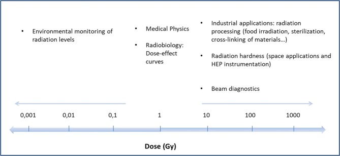
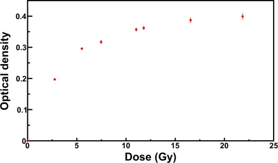
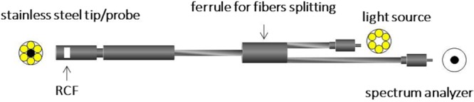
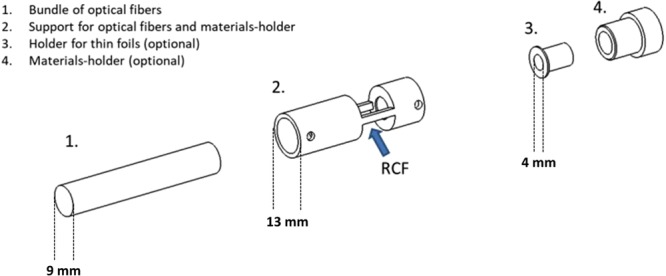
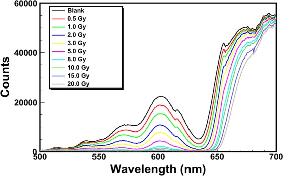
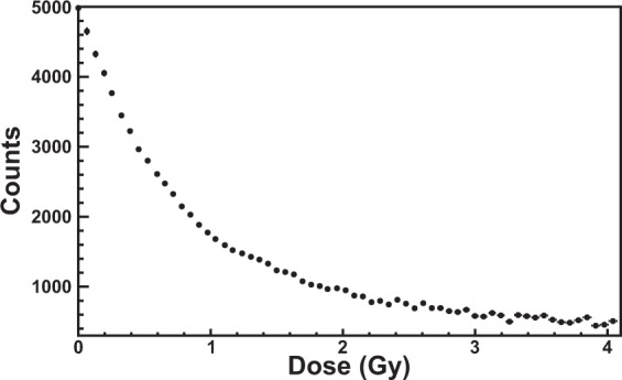
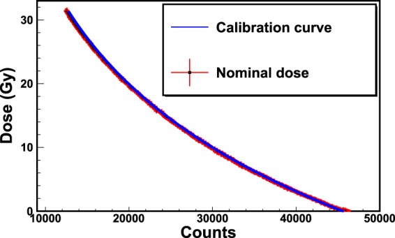
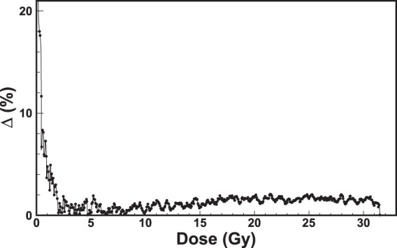
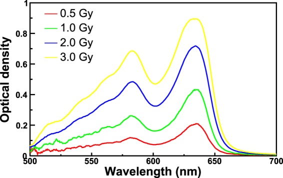
Radiochromic film dosimetry has been widely employed in most of the applications of radiation physics for over twenty years. This is due to a number of appealing features of radiochromic films, such as reliability, accuracy, ease of use and cost. However, current radiochromic film reading techniques, based on the use of commercial densitometers and scanners, provide values of dose only after the exposure of the films to radiation. In this work, an innovative methodology for the real-time reading of radiochromic films is proposed for some specific applications. The new methodology is based on opto-electronic instrumentation that makes use of an optical fiber probe for the determination of optical changes of the films induced by radiation and allows measurements of dose with high degree of precision and accuracy. Furthermore, it has been demonstrated that the dynamic range of some kinds of films, such as the EBT3 Gafchromic films (intensively used in medical physics), can be extended by more than one order of magnitude. Owing to the numerous advantages with respect to the commonly used reading techniques, a National Patent was filed in January 2018.


Conflict of interest statement

The authors declare no competing interests.

Figures

Figure 1
Figure 1
Applications of the radiation physics in which RCFs are employed.
Figure 2
Figure 2
Calibration of EBT3 Gafchromic films exposed to 1 MeV electron beam. The analysis was performed with the EPSON V800 Scanner.
Figure 3
Figure 3
Experimental apparatus for the real-time reading of RCFs.
Figure 4
Figure 4
Design of mechanical elements for the support of the backscattering material.
Figure 5
Figure 5
Picture of the terminating part of the optical fiber bundle with the RCF (a) and top view (b).
Figure 6
Figure 6
Picture of the Gammacell220 of ISOF-CNR at Bologna. During the operation the optical fiber probe is inside the irradiator. The light source and the spectrometer are remotely controlled.
Figure 7
Figure 7
Spectra of an EBT3 Gafchromic film exposed to 60Co gamma-rays from the HDR-irradiator. The integration-time is set to 1.5 s.
Figure 8
Figure 8
Counts corresponding to the wavelength λlow = 635 nm as a function of the dose. Above 4 Gy this physical quantity is not useful for the calibration (saturation).
Figure 9
Figure 9
Calibration of an EBT3 Gafchromic film exposed to 60Co-gamma rays of the HDR-irradiator. The counts corresponding to the wavelength λlow = 663 nm are used for the calibration. The red curve is the exponential function that best fits the data. The values of χ2/ndf, χ2-probability and the fitting parameters are shown in the inset.
Figure 10
Figure 10
Calibration of an EBT3 Gafchromic film exposed to 60Co-gamma rays of the LDR-irradiator. The counts corresponding to the wavelength λlow = 635 nm are used for the calibration. The red curve is the exponential function that best fits the data. The values of χ2/ndf, χ2, -probability and the fitting parameters are shown in the inset.
Figure 11
Figure 11
Spectra of an XR-QA2 Gafchromic film exposed to 60Co gamma-rays from the LDR-irradiator. The integration-time is set to 630 ms.
Figure 12
Figure 12
Calibration of an XR-QA2 Gafchromic film exposed to 60Co-gamma rays of the LDR-irradiator. The counts corresponding to the wavelength λlow = 635 nm are used for the calibration. The black curve is the exponential function that best fits the data. The zoom of the main plot in the range of dose [0–200 mGy] is shown in the first inset. The values of χ2/ndf, χ2, -probability and the best estimates of the fitting parameters are shown in the second inset.
Figure 13
Figure 13
Validation of the new RCF real-time reader. The plot shows the dose as a function of counts for an exposure of an EBT3 Gafchromic film to the HDR irradiation. The nominal doses (black dots) are compared to the calibrated doses (blue curve). The maximum uncertainty was estimated by computing the percentage difference (Δ %) between calibrated and nominal doses. Figure 14 shows the maximum uncertainty as a function of dose for the values of Fig. 13.
Figure 14
Figure 14
Percentage difference (Δ %) as a function of dose for the values of Fig. 13.
Figure 15
Figure 15
Optical density spectra obtained by evaluating the OD of the spectra of Fig. 7.

References

    1. Williams, M. & Metcalfe, P. Radiochromic film dosimetry and its applications in radiotherapy. In AIP Conference Proceedings, vol. 1345, 75–99 (AIP, 2011).
    1. McLaughlin WL, Chalkley L. Measurement of radiation dose distributions with photochromic materials. Radiol. 1965;84:124–125. doi: 10.1148/84.1.124. - DOI - PubMed
    1. Humphreys JC. Nist high-dose calibration services. Nucl. Instruments Methods Phys. Res. Sect. B: Beam Interactions with Mater. Atoms. 1989;40:1173–1177. doi: 10.1016/0168-583X(89)90565-X. - DOI
    1. McLaughlin WL, et al. Novel radiochromic films for clinical dosimetry. Radiat. protection dosimetry. 1996;66:263–268. doi: 10.1093/oxfordjournals.rpd.a031731. - DOI
    1. Soares CG. Radiochromic film dosimetry. Radiat. measurements. 2006;41:S100–S116. doi: 10.1016/j.radmeas.2007.01.007. - DOI