. 2019 Mar 29;9(1):5388.
doi: 10.1038/s41598-019-41959-8.

Intense bone fluorescence reveals hidden patterns in pumpkin toadlets

Affiliations

Intense bone fluorescence reveals hidden patterns in pumpkin toadlets

Sandra Goutte et al. Sci Rep. .

Abstract

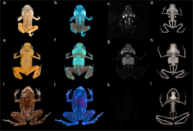
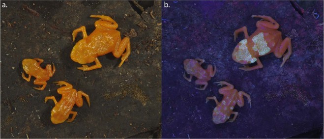
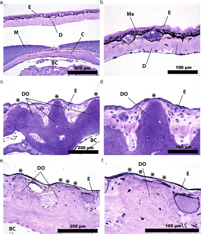
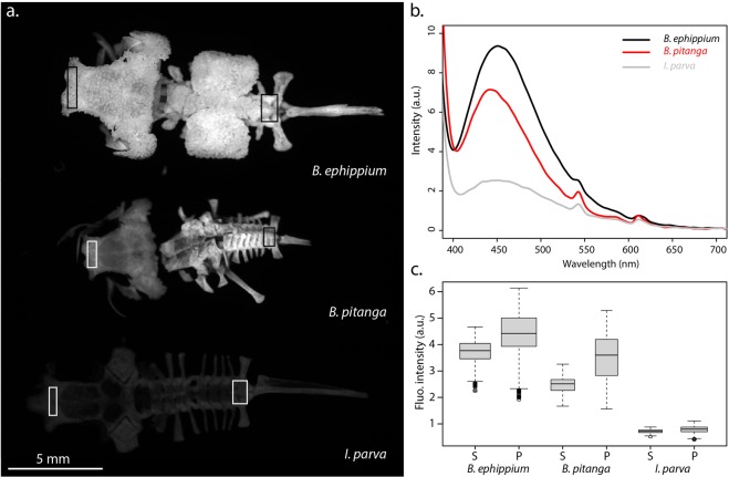
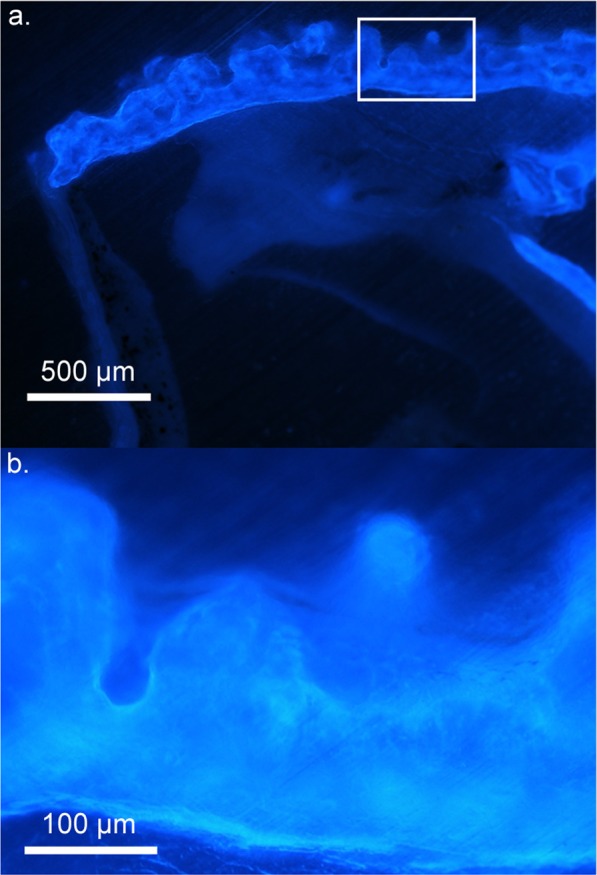
The phenomenon of fluorescence can be used by animals to change effective colouration or patterning, potentially to serve functions including intra- and interspecific signalling. Initially believed to be restricted to marine animals, fluorescent colours are now being described in an increasing number of terrestrial species. Here, we describe unique, highly fluorescent patterns in two species of pumpkin toadlets (Brachycephalus ephippium and B. pitanga). We establish that the origin of the fluorescence lies in the dermal bone of the head and back, visible through a particularly thin skin. By comparing them to those of the closely related species Ischnocnema parva, we demonstrate that pumpkin toadlets' bones are exceptionally fluorescent. We characterize the luminescence properties of the toadlets' bones and discuss the potential function of fluorescent patterns in natural lighting conditions.


Conflict of interest statement

The authors declare no competing interests.

Figures

Figure 1
Figure 1
Fluorescence in pumpkin toadlets. Ethanol-preserved specimens of Brachycephalus pitanga (ac), B. ephippium (eg) and Ischnocnema parva (k), and live Ischnocnema parva (i,j) photographed in natural light (a,e,i) and showing fluorescence under UV illumination using two Fluotest Forte UV (λexcitation centred around 365 nm; b,f,j) and a laboratory UV light source (λexcitation = 365 nm) and an emission filter centred around 472 nm and 30 nm wide, thereby eliminating reflectance of all visible light (c,g,k). Note that the absence of fluorescence in I. parva results in a completely dark image (k). Computerized micro-tomography (µCT) reconstructions (c,h,l) show the correspondence between fluorescent patterns and bone structure in B. pitanga (d), B. ephippium (h) and I. parva (l). Photographs taken by L.C. and S.G. (a,b,e,f,i,j) and P.G., M.T. and S.G. (c,g,k).
Figure 2
Figure 2
Development of fluorescence in Brachycephalus ephippium. Three live individuals of B. ephippium share the same skin colour (a: natural lighting) but present different levels of fluorescence (b; UV illumination using two SANKYO lamps λexcitation = 315–400 nm, no filter), corresponding to the extent of dermal ossification. The smallest individual (juvenile) does not present any fluorescent pattern; the larger juvenile presents several fluorescent points on its back and head; the adult presents the complete pattern, with more obvious fluorescence of the head and back. Photographs taken by S.G. and C.J.
Figure 3
Figure 3
Dermal ossification in pumpkin toadlets. Photomicrographs of transverse histological sections of heads of Brachycephalus hermogenesi (a,b), B. ephippium (c,d) and B. pitanga (e,f). BC: brain cavity, C: cranial bone, D: dermis, DO: dermal ossification, E: epidermis, M: muscle, Me: melanophore. Asterisks indicate points where the fluorescent ossified tissue is visible through the thin epidermis in live specimens. B. hermogenesi lacks dermal ossification and a layer of melanophores is present, in contrast to B. ephippium and B. pitanga.
Figure 4
Figure 4
Bone fluorescence in pumpkin toadlets. (a) Emission of bones of Brachycephalus ephippium, B. pitanga and Ischnocnema parva under UVA lighting (λexcitation = 365 nm) collected in the spectral range where their fluorescence occurs, using a 455–485 nm band-pass filter. The bones represented are the skull, spinal column and urostyle. All were imaged in a single shot to allow direct comparison of their emission intensities. (b) Emission spectra of the skull (1.5 mm diameter area) of the three species under UV lighting (λexcitation = 365 nm), in arbitrary units (a.u.). (c) Boxplots, corresponding to the boxes in (a), comparing fluorescence intensities (in terms of grey-level of single pixels) of the skull (S) and pelvic area (P) of one individual for each of the three species (B. ephippium: n = 1,760 pixels (S), n = 2,331 pixels (P); B. pitanga: n = 1,064 pixels (S), n = 943 pixels (P); I. parva: n = 1,054 pixels (S), n = 1,320 pixels (P)). The boxes represent the first and the third quartiles; the bold line the median; the whiskers the first and ninth deciles; and the points outliers. Quantitative indices are given in Table S1.
Figure 5
Figure 5
Fluorescence distribution within the bone in Brachycephalus ephippium. Photomicrograph of a transverse, non-decalcified section of the dorsal bony plates at 10x (a) and an enlargement of the boxed area at 40x magnification (b). The section is illuminated with UV-A light (λexcitation = 365 nm) and no emission filter was used.

References

    1. Arnold KE, Owens IPF, Marshall NJ. Fluorescent signaling in parrots. Science. 2002;295:92. doi: 10.1126/science.295.5552.92. - DOI - PubMed
    1. Lim MLM, Land MF, Li D. Sex-specific UV and fluorescence signals in jumping spiders. Science. 2007;315:481–481. doi: 10.1126/science.1134254. - DOI - PubMed
    1. Taboada, C. et al. Naturally occurring fluorescence in frogs. Proc. Natl. Acad. Sci. 201701053, 10.1073/pnas.1701053114 (2017). - PMC - PubMed
    1. Prötzel D, et al. Widespread bone-based fluorescence in chameleons. Sci. Rep. 2018;8:698. doi: 10.1038/s41598-017-19070-7. - DOI - PMC - PubMed
    1. Gruber DF, et al. Biofluorescence in catsharks (Scyliorhinidae): fundamental description and relevance for elasmobranch visual ecology. Sci. Rep. 2016;6:24751. doi: 10.1038/srep24751. - DOI - PMC - PubMed

Publication types