. 2019 Jul 9;47(12):e69.
doi: 10.1093/nar/gkz222.

PIXUL-ChIP: integrated high-throughput sample preparation and analytical platform for epigenetic studies

Affiliations

PIXUL-ChIP: integrated high-throughput sample preparation and analytical platform for epigenetic studies

Karol Bomsztyk et al. Nucleic Acids Res. .

Abstract

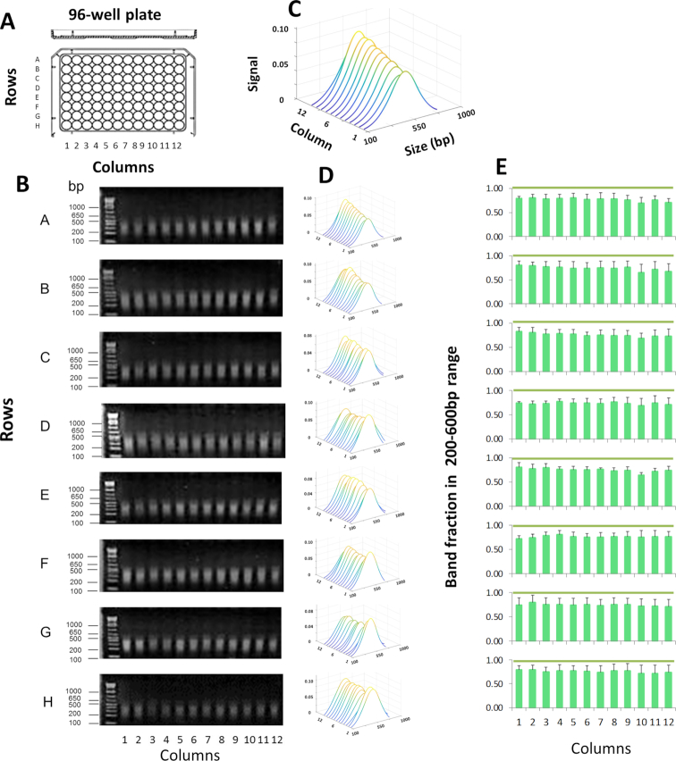
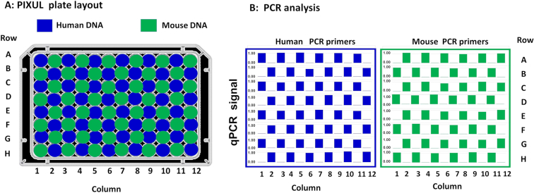
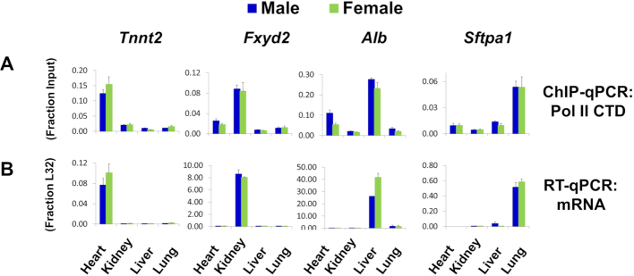
Chromatin immunoprecipitation (ChIP) is the most widely used approach for identification of genome-associated proteins and their modifications. We have previously introduced a microplate-based ChIP platform, Matrix ChIP, where the entire ChIP procedure is done on the same plate without sample transfers. Compared to conventional ChIP protocols, the Matrix ChIP assay is faster and has increased throughput. However, even with microplate ChIP assays, sample preparation and chromatin fragmentation (which is required to map genomic locations) remains a major bottleneck. We have developed a novel technology (termed 'PIXUL') utilizing an array of ultrasound transducers for simultaneous shearing of samples in standard 96-well microplates. We integrated PIXUL with Matrix ChIP ('PIXUL-ChIP'), that allows for fast, reproducible, low-cost and high-throughput sample preparation and ChIP analysis of 96 samples (cell culture or tissues) in one day. Further, we demonstrated that chromatin prepared using PIXUL can be used in an existing ChIP-seq workflow. Thus, the high-throughput capacity of PIXUL-ChIP provides the means to carry out ChIP-qPCR or ChIP-seq experiments involving dozens of samples. Given the complexity of epigenetic processes, the use of PIXUL-ChIP will advance our understanding of these processes in health and disease, as well as facilitate screening of epigenetic drugs.


Figures

Figure 1.
Figure 1.
PIXUL shearing of DNA in 96-well plates. (A) Shearing was performed in 96-well plates (with each well containing salmon DNA at 100 ng/μl in 100μl volume/well) for a total treatment time of 36 min per each plate. (B) Agarose gel electrophoresis of DNA fragments, gels were stained with ethidium bromide. DNA ladder was run in the first lane of each gel. Numbers to the left of the gels show sizes of selected ladder bands in base pair (bp). (C) An example to illustrate a waterfall plot (MATLAB) with annotated axis. Image software was used to analyze stained DNA bands (Methods). Results represent best-fit curves in sequential order of samples from PIXUL plate column wells 1 to 12. X- axis; band size in base pair (Size (bp)). Y-axis; sample from a well of a given column (columns 1–12). Z-axis; relative signal intensity of DNA bands for given plate well (Signal). (D) Waterfall plots for each plate row (rows A through H). (E) Graphs represent band fraction in the 200–600 bp range from each one of the 96 wells (mean ± SDEV, n = 3 experiments). These results demonstrate consistent DNA shearing across all wells of a 96-well plate.
Figure 2.
Figure 2.
PIXUL shearing of chromatin directly in 96-well plate cell cultures. (A) HCT116 cell cultures grown in 96-well plates were crosslinked directly in plates followed by glycine quenching. After PBS wash, shearing buffer was added. Plates were then sealed and were treated in PIXUL (total time 36 min per plate). After digestion with proteinase K and reversal of crosslinking, sheared DNA fragments were resolved by agarose gel electrophoresis. (B) Agarose, gels were stained with ethidium bromide. DNA ladder was run in the first lane of each gel. Numbers to the left of the gels show sizes of selected ladder bands in base pair (bp). (C) An example to illustrate a waterfall plot (MATLAB) with annotated axis. Image software was used to analyze stained DNA bands (Methods). Results are shown as waterfall plots (MATLAB) of best-fit curves in sequential order of samples from culture plate column well 1 to 12. X- axis; band size in base pair (size (bp)). Y-axis; sample from a well of a given column (columns 1 through 12). Z-axis; relative signal intensity of bands for given plate well (Signal). (D) Waterfall plots for each plate row (rows A through H). (D) Waterfall plots for each plate row. (E) Graphs represent band fraction in the 200–600 bp range from each one of the 96 wells (mean ± SDEV, n = 4 experiments). These results show that a 96-well plate culture can be directly sonicated with PIXUL, avoiding the sample transfer step and yielding consistent chromatin fragmentation across all 96 wells.
Figure 3.
Figure 3.
Across 96-well plate contamination test. (A) Human and mouse genomic DNA (10 ng/μl in 100 μl volume) were loaded into 96-well plate in a checkerboard layout. After sealing with a film adhesive, plate was treated with PIXUL (18 min per plate), and DNA in each well was assessed using human (EGR1) and mouse (Tnfa) primers in qPCR. (B) Results of qPCR analysis with human (left panel) and mouse (right panel) primers for each one of the 96-wells (rows A–H and columns 1–12). Bars (blue or green) in the graphs show relative human and mouse DNA concentrations (each well/average non-zero concentration across the entire plate- scale shown 0.0–1.0). These results demonstrate that there is no detectable (not different from 0.0) cross-contamination across wells.
Figure 4.
Figure 4.
PIXUL-ChIP-qPCR analysis of Pol II recruitment kinetics to the EGR1 locus in serum-treated 96-well HCT116 culture. Serum-deprived HCT116 96-well cultures were treated with serum for 0, 5, 15 and 30 min. Cells were crosslinked directly in the 96-well plate, quenched with glycine, and washed with PBS. PBS was then replaced with shearing buffer, and the plate was treated with PIXUL. Sheared chromatin was used in Matrix ChIP-qPCR analysis of Pol II at the EGR1 gene. (A) Layout of the serum time-course treatment experiment. (B, C) Pol II ChIP-qPCR analysis at the EGR1 (B) and UBE2b (C) loci presented as fraction of input. Graphs show mean ± SEM (n = 4) of combined ChIP-qPCR as shown (n = 4 wells/each time point). Gray/blue boxes above the graphs correspond to colors of the plate quadrants in (A). Cartoons of the EGR1 and UBE2b genes and location of the PCR primers (colored boxes) are shown below. These results show that cells grown in 96-well plate can be treated with an inducing agent (here, serum) and sonicated directly on the culture plate in PIXUL (no sample transfer), and then sheared chromatin aliquots analyzed in microplate ChIP-qPCR, yielding reproducible results of all 96 samples in one day.
Figure 5.
Figure 5.
PIXUL-ChIP-qPCR analysis of Pol II kinetics of recruitment to inducible loci in response to serum- and TPA-treatment of 96-well HCT116 and HEK293 cell cultures. Serum-deprived HCT116 and HEK293 cultures in the same 96-well plate were treated with either 10% serum or 100 nM TPA for 5, 15, 30 min and 1, 2, 4, 6, 18, 24 and 48 h. Cells were crosslinked, plates sealed and treated with PIXUL as in Figure 2. Sheared chromatin was used in microplate ChIP analysis of Pol II density at EGR1 and NR4A3 genes. (A) 96-well plate culture layout of the serum and TPA time-course treatment experiment. (B) Graphs of ChIP-qPCR results showing Pol II density (as a fraction of input), mean ± SEM (n = 2) of respective cell lines, treatments (serum; green, TPA;blue) and harvested at indicated time points. (C) Gene cartoons and position of PCR primers. These data show that different cells can be cultured on the same 96-well plate, treated with different agents at various times, and then sheared directly in PIXUL and analyzed by ChIP-qPCR yielding results for all 96 samples in one day.
Figure 6.
Figure 6.
PIXUL-ChIP-qPCR analysis of Pol II and epigenetic modifications at the OCT4 (POU5F1) locus in hESC Elf1 cells. Human embryonic stem cells (hESC Elf1) were cultured in 96-well plates as naive (2i + hLif + FGF2 + Igf1) or as primed (TeSR+FGF2) on Matrigel for either one or two passages. (2i- two inhibitors: PD0325901 MEKi and CHIR-99021GSK3i). One and two passages in TeSR represent cells transitioning to primed. These cells were plated at 10 000 cells/well on Matrigel in 96-well plates with Rho kinase (ROCK) inhibitor present for the first 24 h of culture to improve survival (32). Half of the plate was used to extract RNA for RT-qPCR (normalized to L32 mRNA) (A) and the other half (chromatin) was crosslinked, sheared in PIXUL, and subjected to Matrix ChIP analysis (expressed as a fraction of input) (B) as in Figure 3. Statistical differences between two means (P value) are shown by the size of the solid circles: P < 0.05 for small circle, P < 0.01 for large circle, and no circle indicating the differences are not statistically significant (7). These results are consistent with previous observations and as such demonstrated that PIXUL-ChIP-qPCR platform could be used for high-throughput experiments and drug screening.
Figure 7.
Figure 7.
Matrix ChIP analysis of chromatin prepared from 96-well HEK293 cultures using PIXUL and Bioruptor. Serum-deprived HEK293 96-well cultures were treated with serum for 0, 5, 15 and 30 min. Cells were crosslinked directly in the 96-well plate, harvested manually from Row A (12 samples) and transferred to twelve 0.5 ml tubes for ultrasound treatment in the Bioruptor (45 min). The rest of the plate was sealed and sonicated in PIXUL (26 min treatment). (A) comparison of sheared chromatin fragments obtained with Bioruptor versus PIXUL analyzed by agarose gel electrophoresis, Ethidium bromide stained gels are shown, sizes (bp) of DNA ladder fragments (first lane) are shown to the left. Sonicated fragments were analyzed by image analysis software (Methods), results are displayed as waterfall plots in sequential order of samples run from lane 1 to 12. X- axis; band size in base pair (bp). Y-axis; sample from a given lane. Z-axis; relative signal intensity of bands. (B) Mean fragment size (each blue dot) of sheared chromatin obtained with Bioruptor and PIXUL. (C) Sheared chromatin samples from both PIXUL and Bioruptor were analyzed simultaneously by Matrix ChIP using antibodies to Pol II, pB-Raf, pErk and H3K27m3. ChIPed DNA was analyzed by real-time PCR using indicated primers. Results show mean ± SEM (n = 3 replicates for each time point for each instrument). This comparison demonstrates that sample transfer causes significant sample losses, which may in part account for greater variability. There are lower Matrix ChIP signals from chromatin prepared with Bioruptor compared to PIXUL.
Figure 8.
Figure 8.
Matrix ChIP analysis of chromatin prepared from 96-well HCT116 cultures using either PIXUL or Covaris LE220. Serum-deprived HCT116 cell 96-well plate cultures were treated with serum for 0, 5, 15 and 30 min. Cells were cross-linked directly in the 96-well plate. With Covaris LE220 shearing harvesting cells from one well of a 96-well plate yielded insufficient amounts of chromatin to generate reproducible ChIP results. Thus, with this instrument, for each time point cells harvested from three wells of a 96-well plate were combined into one sample and transferred to either Covaris microplate tubes or Covaris microplate. Each time point was done in duplicate, for a total of eight samples. The rest of the 96-well plate was sealed and sonicated in PIXUL (18 min treatment). (A–C) Agarose gel electrophoresis analysis of chromatin fragments sonicated using Covaris LE220 microplate tubes (A), Covaris LE220 plate (B) or PIXUL (C). Sheared fragments were analyzed by image analysis software (Methods), results are displayed as waterfall plots in sequential order of samples run from lane 1 to 8 as in Figure 1. X- axis; band size in base pair (bp). Y-axis; sample from a given lane. Z-axis; relative signal intensity of bands. Numbers above the plots show average fragment size ±SEM for all eight samples. (D) Sizes of chromatin samples (A-C) sonicated by either Covaris tubes, Covaris plate or PIXUL. (E) Sheared chromatin samples prepared using either Covaris tubes (cells from 3 wells combined into one sample) and PIXUL (single well per sample) were analyzed simultaneously by Matrix ChIP using antibodies to Pol II, H3K9Ac, H3K27Ac, H3K27m3, H3K36m3 and CTCF. ChIP DNA was analyzed by qPCR using indicated primers. Results show fraction of input, mean±SEM (n = 3 replicates for each time point for each instrument). (F) Comparison of Pol II and CTCF ChIP signals at known binding and respective distal sites using Covaris versus PIXUL sonicated chromatin (yellow circles below graphs show P < 0.05). (G) EGR1 gene cartoon and position of PCR primers. This comparison demonstrates that sonication of chromatin with PIXUL is more consistent and yields smaller fragments compared to Covaris; in particular the first position (A1) in their plate yields considerably smaller fragment than the other wells. Combing cells from three wells of a 96-well plate for Covaris sonication generates chromatin yielding similar ChIP results to those obtained using cells from one well of a 96-well plate treated with PIXUL. The ChIP background signal is lower with PIXUL compared to Covaris.
Figure 9.
Figure 9.
PIXUL-ChIP analysis of Pol II occupancy in mouse heart, kidney, liver, and lung. Flash frozen heart, kidney, liver and lung samples from male and female mice were cross-linked and then sonicated in microplates using PIXUL. (A) PIXUL-sheared chromatin samples were simultaneously analyzed for Pol II levels at indicated organ-specific genes using Matrix ChIP. ChIP DNA was analyzed by qPCR expressed as fraction of input. Data represent mean ± SEM (n = 3 mice) expressed as a fraction of input. (B) RNA isolated from the same frozen organs as in A was used in RT-qPCR with primers to indicated genes. Data represent mean ± SEM (n = 3 mice) expressed as a ratio to the transcript levels of housekeeping ribosomal protein gene, L32. These results demonstrate that PIXUL-ChIP can be used to analyze multiple samples from several organs on the same plate.
Figure 10.
Figure 10.
PIXUL-ChIP-seq results and comparison to ENCODE datasets. HCT116 cells were grown to the density of ∼200 000 cells per well, cross-linked, and sonicated using 96-well PIXUL. ChIP was performed and libraries were generated from a single PIXUL well using Active Motif's Low Cell ChIP-Seq Kit. Libraries were sequenced on a NextSeq 500 (Methods). (A) Number of peaks in PIXUL-ChIP-seq and ENCODE datasets, and percentage of PIXUL-ChIP-seq peaks that are also detected in ENCODE. (B) PIXUL-ChIP-seq (white background) and ENCODE (gray background) genome browser snapshot of a region around the EGR1 locus occupied by CTCF, H3K9Ac, H3K27Ac, H3K4m1, H3K4m3, H3K36m3 and H3K27m3. The data demonstrate good agreement between PIXUL-ChIP-seq (which was done in ∼200,000 cells) compared to ENCODE (which used >106 cells). (C–H) To verify that genes marked by PIXUL-ChIP-seq peaks show expected expression patterns (genes with repressive marks have lower expression, genes with active marks show higher expression), HCT116 RNA-seq data were downloaded from Sanger Institute Genomics of Drug Sensitivity in Cancer (GDSC) website (https://www.cancerrxgene.org/). Expression distribution was plotted of genes with histone marks at the transcription start sites (TSS) and those without. Genes marked with active histone marks around TSS have a mean expression of 32 (25) Fragments Per Kilobase Million (FPKM), while genes without active histone marks (or with repressive mark H3K27m3) are expressed < 1FPKM – resulting in the bimodal distribution. (C) Expression distribution for genes with H3K4m1 PIXUL-ChIP-seq peaks (orange) around TSS and genes without H3K4m1 peaks (blue). (D) Expression distribution for genes with H3K4m3 peaks around TSS and genes without H3K4m3 peaks. (E) Expression distribution for genes with H3K9Ac peaks around TSS and genes without H3K9Ac peaks. (F) Expression distribution for genes with H3K36m3 peaks around TSS and genes without H3K36m3 peaks. (G) Expression distribution for genes with H3K27Ac peaks within gene body or around TSS and genes without H3K27Ac peaks. (H) Expression distribution for genes with H3K27m3 peaks within gene body or around TSS and genes without H3K27me3 peaks.

References

    1. Orlando V., Strutt H., Paro R.. Analysis of chromatin structure by in vivo formaldehyde cross-linking. Methods. 1997; 11:205–214. - PubMed
    1. Solomon M.J., Varshavsky A.. Formaldehyde-mediated DNA-protein crosslinking: a probe for in vivo chromatin structures. Proc. Natl. Acad. Sci. U.S.A. 1985; 82:6470–6474. - PMC - PubMed
    1. O’Neill L.P., Turner B.M.. Immunoprecipitation of chromatin. Methods Enzymol. 1996; 274:189–197. - PubMed
    1. Huebert D.J., Kamal M., O’Donovan A., Bernstein B.E.. Genome-wide analysis of histone modifications by ChIP-on-chip. Methods. 2006; 40:365–369. - PubMed
    1. Heintzman N.D., Stuart R.K., Hon G., Fu Y., Ching C.W., Hawkins R.D., Barrera L.O., Van Calcar S., Qu C., Ching K.A. et al. .. Distinct and predictive chromatin signatures of transcriptional promoters and enhancers in the human genome. Nat. Genet. 2007; 39:311–318. - PubMed

Publication types