Review
. 2019 Mar 29;10(1):42.
doi: 10.1186/s13244-019-0728-4.

The imaging appearances of various pericardial disorders

Affiliations
Review

The imaging appearances of various pericardial disorders

Emre Ünal et al. Insights Imaging. .

Abstract

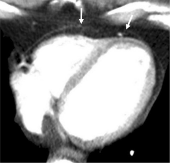
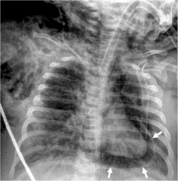
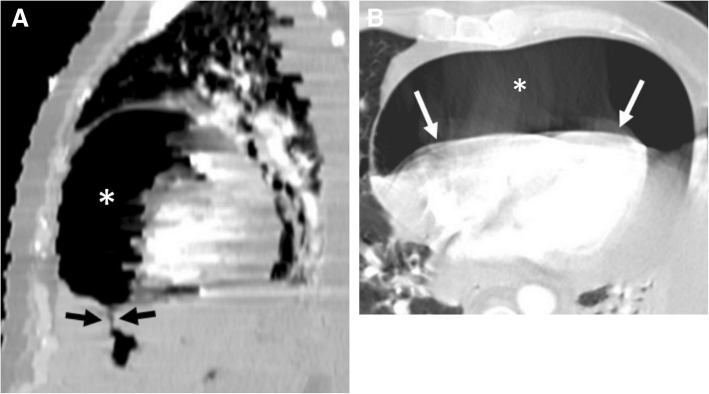
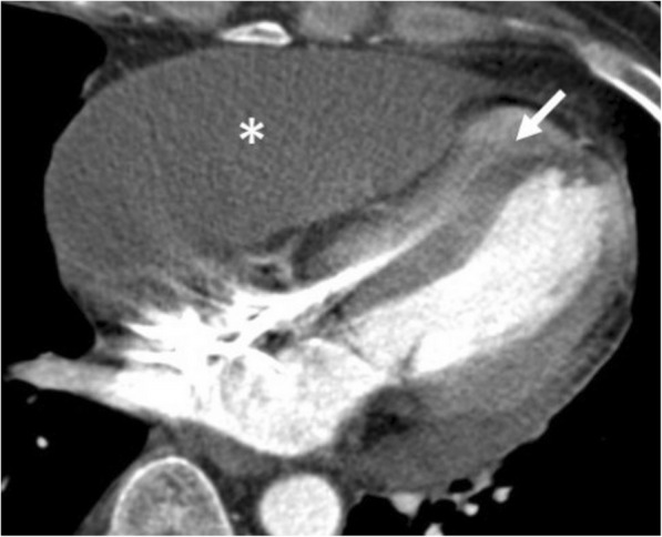
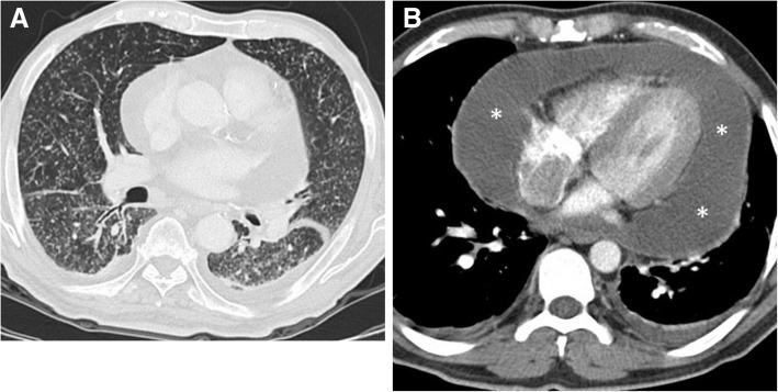
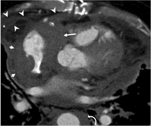
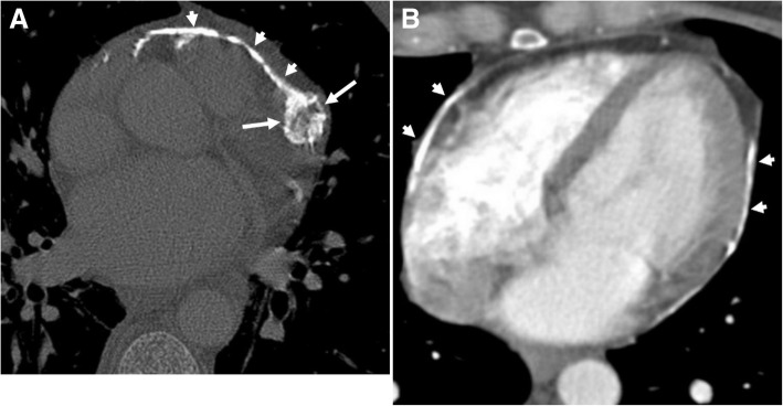
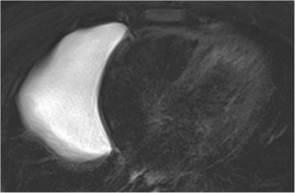
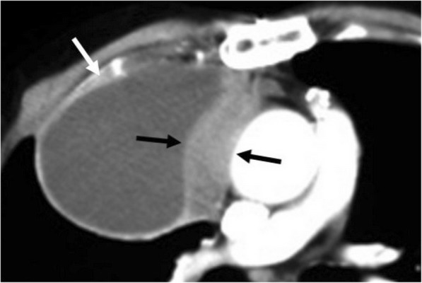
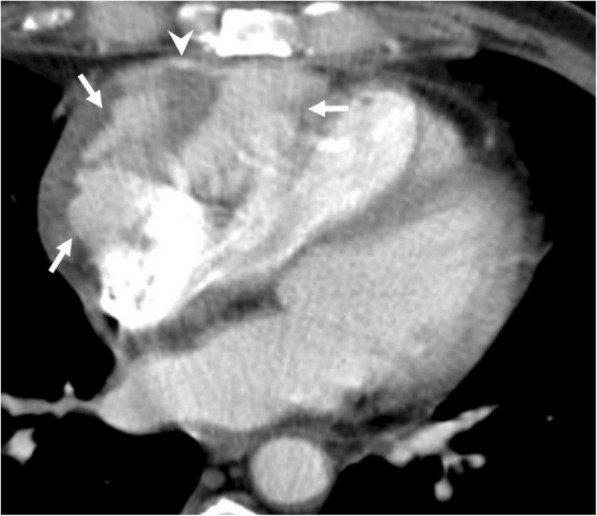
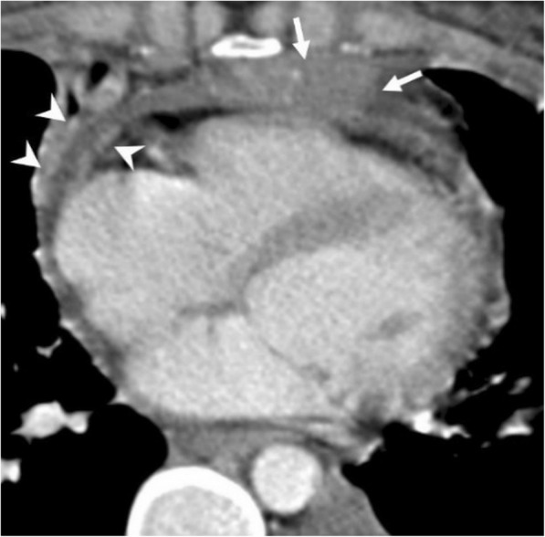
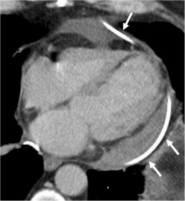
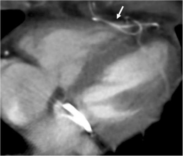
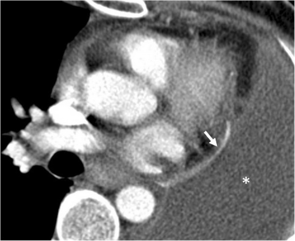
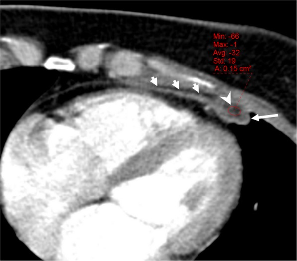
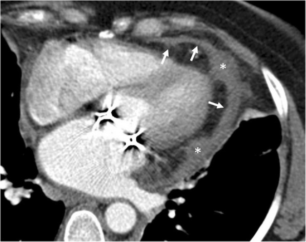
The pericardium could be involved in a variety of clinical disorders. The imaging findings are not specific for an individual pathology in most of the cases; however, patient's clinical history may guide radiologist to a definitive diagnosis. Congenital absence of the pericardium could be recognized with the imaging appearance of interposed lung tissue between the main pulmonary artery and aorta. Pericardial effusion is a non-specific condition that may occur due to inflammatory, infectious, and neoplastic disorders. Cardiac tamponade may occur in case of massive or rapid accumulation of fluid in the pericardial sac. Pericardial calcification is a common and easily identified entity on a computed tomography (CT) scan. Presence of calcification and/or fibrosis may result in pericardial constriction. Nevertheless, the pulsation of an adjacent coronary artery may prevent calcification formation in a focal area and consequently may result in pericardial diverticulum containing epicardial fat and coronary artery. The imaging findings encountered in patients with pericardial hydatid disease and Erdheim-Chester disease may mimic those of pericardial neoplasia. Pericardial adhesions and pedicled fat flaps may cause confusion on a CT scan in the post-surgical period following cardiac surgery. Pericardial fat necrosis can be diagnosed by CT in patients with chest pain. The radiologists should be familiar with the medical devices placed in pericardial space for certain individual indications. A pericardial patch and temporary epicardial pacemaker wires could be identified on a CT scan.

Keywords: Absence of the pericardium; Gastropericardial fistula; Hydatid disease; Pericardial diverticulum; Pericardial mesothelioma; Pericardial metastases; Pericardial patch.


Conflict of interest statement

Competing interests

The authors declare that they have no competing interests.

Publisher’s Note

Springer Nature remains neutral with regard to jurisdictional claims in published maps and institutional affiliations.

Figures

Fig. 1
Fig. 1
A 51-year-old man. Axial contrast-enhanced CT (a) and T2-weighted MR images (b) demonstrate levorotation (curved arrows) of the heart and lack of pericardial continuity (arrows). c Axial CT image of a different patient with congenital absence of the pericardium demonstrates a prominent main pulmonary artery (PA). Interposed lung tissue (arrow) between the main pulmonary artery and the ascending aorta (AA)
Fig. 2
Fig. 2
A 68-year-old man with previous coronary artery bypass surgery. Axial contrast-enhanced CT image demonstrates the lack of pericardial continuity (arrows) due to surgery
Fig. 3
Fig. 3
A 3-month-old girl with pneumonia and sepsis developed pneumopericardium (arrows) under endotracheal intubation at insensitive care unit
Fig. 4
Fig. 4
A 51-year-old man with pathologically proven gastric lymphoma developed sudden onset of dyspnea and cardiac arrhythmia. Contrast-enhanced CT in the sagittal (a) and axial (b) planes demonstrates a gastropericardial fistula (black arrows) complicated with massive pneumopericardium (asterisks). Significant compressive effect to the heart is also noted (white arrows)
Fig. 5
Fig. 5
A 61-year-old man with right heart failure symptoms underwent to CT scan to rule out the presence of pulmonary artery embolism. Axial contrast-enhanced CT image shows significantly compressed right ventricle (arrow) due to the loculated pericardial effusion (asterisk)
Fig. 6
Fig. 6
A 86-year-old woman with acute onset of pain in the chest. Axial contrast-enhanced CT image reveals hemopericardium (arrowheads) and intramural hematoma of the ascending aorta (asterisks)
Fig. 7
Fig. 7
A 52-year-old woman with breast cancer receiving radiotherapy for treatment. Axial contrast-enhanced CT image reveals pericardial effusion with increased density compatible with hemopericardium (arrowheads)
Fig. 8
Fig. 8
A 41-year-old man with newly diagnosed pulmonary tuberculosis. a Axial contrast-enhanced CT image at lung window reveals miliary pattern of pulmonary involvement. b Large amount of pericardial effusion (asterisks) is also noted. A culture test was positive for tuberculosis following the US-guided pericardiocentesis
Fig. 9
Fig. 9
A 61-year-old man with Erdheim-Chester disease. Axial reformatted contrast-enhanced CT image demonstrates lobulated soft tissue densities with pseudo-tumor appearance involving the right atrium (short arrows), right atrioventricular groove (long arrow), and the aortic wall (curved arrow). Pericardial (asterisk) effusion and thickening (arrowheads) are also seen
Fig. 10
Fig. 10
a, b Axial CT images of two different patients demonstrate the nodular (long arrows) and linear (short arrows) areas of calcification through the course of pericardium
Fig. 11
Fig. 11
A 35-year-old man underwent to CT scan due to decreased cardiac output. a Axial CT image reveals entrapped heart appearance (arrows) with diffuse pericardial calcification which is compatible with constrictive pericarditis. b Axial contrast-enhanced CT image following surgery demonstrates diminished compressive effect to the right side of the heart; however, residual compressive effect to the left ventricle (arrow) was still evident despite surgery
Fig. 12
Fig. 12
62-year-old woman with echocardiography findings suggestive of restrictive cardiomyopathy or constrictive pericarditis. Contrast-enhanced CT images on reformatted coronal (a) and axial plane (b) demonstrate linear pattern of extensive pericardial calcification. A pericardial diverticulum (short arrows) containing epicardial fat, coronary artery (arrowheads) and lateral wall of the right ventricle (long arrow) is noted
Fig. 13
Fig. 13
A 40-year-old man with incidentally detected pericardial cyst. Axial fat-suppressed T2-weighted MR image demonstrates hyperintense cystic mass in the right anterior cardiophrenic angle compatible with a pericardial cyst
Fig. 14
Fig. 14
A 45-year-old man with pathologically proven pericardial mesothelioma. Axial contrast-enhanced CT image shows a heterogeneous pericardial mass with solid (black arrows) and cystic (white arrow) components. Note that there is a lack of fat tissue between the ascending aorta and the mass raising suspicion for perivascular invasion
Fig. 15
Fig. 15
A 59-year-old man with pathologically proven primary cardiac angiosarcoma. Axial contrast-enhanced CT image reveals a highly vascular mass (arrows) invading the right atrioventricular groove, right atrium, and ventricle. Pericardial involvement (arrowhead) is also noted
Fig. 16
Fig. 16
A 26-year-old woman with pathologically proven mediastinal lymphoma. Axial contrast-enhanced CT image demonstrates the inferior aspect of the mediastinal mass (arrows) and pericardial nodular areas of contrast enhancement and thickening suggestive of pericardial involvement (arrowheads)
Fig. 17
Fig. 17
A 28-year-old woman with pathologically proven metastatic osteosarcoma. Axial contrast-enhanced CT image reveals a lobulated hypodense mass (asterisks) invading the anterior aspect of the right ventricle and pericardium
Fig. 18
Fig. 18
A 48-year-old woman with pathologically proven renal cell carcinoma. Axial contrast-enhanced CT image demonstrates the diffuse irregular thickening and contrast enhancement of the pericardium (arrows) compatible with pericardial metastasis
Fig. 19
Fig. 19
A 55-year-old woman with pathologically proven squamous cell carcinoma metastases from unknown origin. a Axial contrast-enhanced CT image demonstrates the nodular areas of pericardial thickening (arrows). b PET-CT scan reveals increased FDG uptake (arrows) at the same areas
Fig. 20
Fig. 20
A 86-year-old man with hydatid disease. Axial contrast-enhanced CT image reveals type 3 hydatid cyst containing multiple daughter cysts (white asterisks). Significantly compressed left atrium (black asterisk) due to hydatid cyst abutting the pericardium
Fig. 21
Fig. 21
A 80-year-old man with echocardiographic findings suggestive of restrictive physiology due to pericardial effusion. Axial contrast-enhanced CT image demonstrates a drainage catheter coursing within pericardial space (arrows)
Fig. 22
Fig. 22
A 45-year-old woman recently underwent mitral valve replacement surgery. Axial contrast-enhanced CT image reveals temporary epicardial pacemaker wires (arrow)
Fig. 23
Fig. 23
A 50-year-old man with lung cancer. Axial contrast-enhanced CT image demonstrates a linear hyperdense pericardial patch (arrow). Large amount of fluid (asterisk) in left pneumonectomy cavity is also noted
Fig. 24
Fig. 24
A 46-year-old woman with a 5-day history of chest pain and shortness of breath. Axial contrast-enhanced CT image demonstrates the fat-containing (arrowhead) soft tissue thickness (long arrow) and associated pericardial thickening (short arrows) compatible with pericardial/epipericardial fat necrosis. The CT findings resolved after conservative treatment
Fig. 25
Fig. 25
A 49-year-old woman with a previous history of mitral valve replacement surgery. Axial contrast-enhanced CT image shows a pericardial effusion (arrows) with irregular and nodular margins (asterisks). Post-surgical adhesions can mimic imaging signs encountered in pericardial metastasis
Fig. 26
Fig. 26
A 65-year-old man with a previous history of surgery for constrictive pericarditis developed chronic sternal osteomyelitis on the follow-up period. Axial contrast-enhanced CT image demonstrates a pedicled fat flap (arrows) placed to maintain hemostasis and infection control. Pericardial thickening (arrowheads) is also noted
Fig. 27
Fig. 27
A 41-year-old war veteran. Axial contrast-enhanced CT image demonstrates a shrapnel fragment abutting the pericardium

References

    1. Wongwaisayawan S, Suwannanon R, Sawatmongkorngul S, Kaewlai R. Emergency thoracic US: the essentials. Radiographics. 2016;36:640–659. doi: 10.1148/rg.2016150064. - DOI - PubMed
    1. Palmer SL, Kelly PD, Schenkel FA, Barr ML. CT-guided tube pericardiostomy: a safe and effective technique in the management of postsurgical pericardial effusion. AJR Am J Roentgenol. 2009;193:W314–W320. doi: 10.2214/AJR.08.1834. - DOI - PubMed
    1. Sun JS, Park KJ, Kang DK. CT findings in patients with pericardial effusion: differentiation of malignant and benign disease. AJR Am J Roentgenol. 2010;194:W489–W494. doi: 10.2214/AJR.09.2599. - DOI - PubMed
    1. Giassi Kde S, Costa AN, Bachion GH, et al. Epipericardial fat necrosis: an underdiagnosed condition. Br J Radiol. 2014;87:20140118. doi: 10.1259/bjr.20140118. - DOI - PMC - PubMed
    1. Rajiah P, Kanne JP. Computed tomography of the pericardium and pericardial disease. J Cardiovasc Comput Tomogr. 2010;4:3–18. doi: 10.1016/j.jcct.2010.01.004. - DOI - PubMed

LinkOut - more resources