. 2020 Mar;23(1):45-53.
doi: 10.1007/s40477-019-00376-3. Epub 2019 Mar 29.

Utility of a gel stand-off pad in the detection of Doppler signal on focal nodular lesions of the skin

Affiliations

Utility of a gel stand-off pad in the detection of Doppler signal on focal nodular lesions of the skin

Antonio Corvino et al. J Ultrasound. 2020 Mar.

Abstract

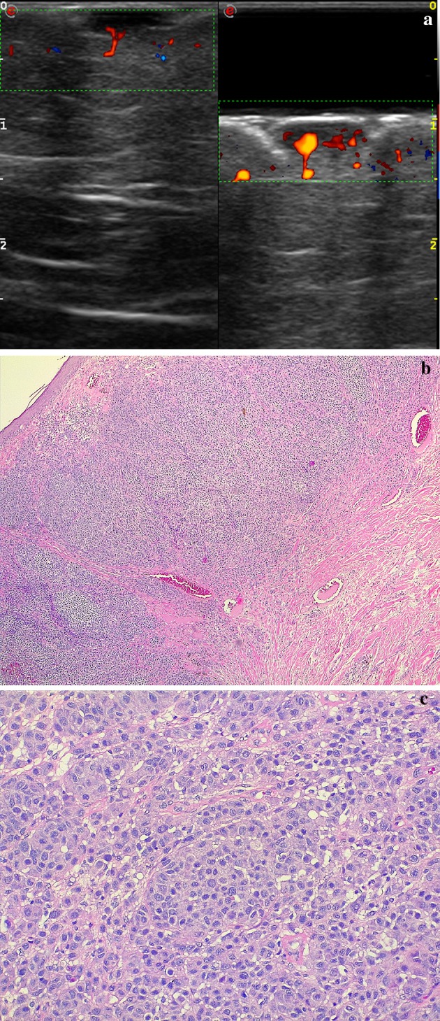
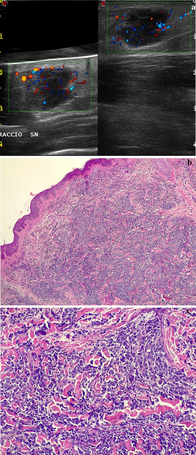
Purpose: Gel pad is an aqueous, flexible, easy available, disposable spacer used for the ultrasound (US) scan of superficial or difficult-to-visualize areas. In clinical practice, it is widely used in B-mode US approach of superficial lesions but, to date, no data have been provided as to its efficacy in the Doppler detection of superficial flows. The aim of our study was to demonstrate the role of stand-off gel pad in the detection of the otherwise-missed peri- or intra-lesional flow signals on Doppler imaging.

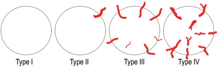
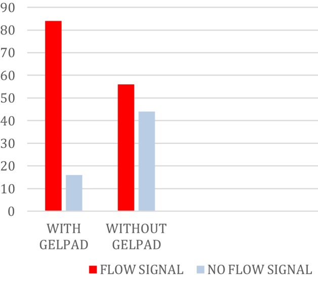
Materials and methods: A total of 100 superficial lesions undergone to an US evaluation using a 7.5-12-MHz linear probe were evaluated prospectively with and without interposition of a gel stand-off pad to detect the presence or absence of vascularization and to classify the vascular pattern.

Results: Peri- or intra-lesional flow was demonstrated in 56% of cases without and in 84% of cases with interposition of a gel stand-off pad; moreover, a statistically significant difference (p value < 0.001) was observed at Chi-square test in the identification of the flow pattern between the use and no use of the pad.

Conclusions: The use of a gel stand-off pad allows the detection of otherwise-missed peri- or intra-lesional flow signals on Doppler imaging, increasing the diagnostic role of this technique in differential diagnosis of superficial lesions.

Keywords: Doppler techniques; Gel stand-off pad; Melanoma; Skin lesions; Skin ultrasound.


Conflict of interest statement

The authors declare that they have no conflict of interest.

Figures

Fig. 1
Fig. 1
Parker Laboratories, Inc (Fairfield, NJ) produces Aquaflex, an aqueous, flexible, easy available disposable, ultrasound gel pad that measures 9 cm in diameter and 2 cm in thickness for the US scan of superficial and difficult-to-visualize areas, which we used in our study
Fig. 2
Fig. 2
The four vascular patterns observed in tumours lesions: avascular (type I), hypovascular with a single vascular pole (type II), hypervascular with multiple peripheral poles (type III), and hypervascular with internal vessels (type IV)
Fig. 3
Fig. 3
Post-surgical back cutaneous melanoma recurrence. a Directional PD technique, split-screen mode (left without spacer, right with spacer). More flow signals can be detected around and within the cutaneous nodule by using a spacer, which allows to better focus the beam superficially. b, c Photomicrographs—hematoxylin-eosin stain, magnification 4x (b) and 20x (c): proliferation of atypical melanocytes arising from the epidermis and infiltrating the dermis and characterized by nests and sheets of cells epithelioid and spindle-shaped with a high nuclear-cytoplasmic ratio, nuclear grooves and pseudoinclusions, large eosinophilic nucleoli and abundant mitotic figures. The immunehistochemistry cytoplasmic positivity for HMB-45 and MART-1 confirmed the diagnosis of melanoma
Fig. 4
Fig. 4
Arm merkel cell carcinoma. a Directional power-Doppler technique. Use of a gel pad to adequately depict the hypoechoic, bulging nodule at US and PD: more peri- and intranodular flow signals are seen using the spacer (left) than without any spacing (right), especially in the uppermost aspect of the nodule. b, c Photomicrographs—hematoxylin-eosin stain, magnification 4x (b) and 20x (c): overgrowth of cells uniform in size arranged in solid sheets and compact nests, which replaced the dermis and subcutaneous tissue without affecting the epidermis. The cytoplasm was scanty but visible as a thin acidophilic rim; the nuclei were round and vesicular with a fine granular chromatin and multiple nucleoli. Mitotic figures and fragmented nuclei were plentiful. The immunohistochemistry cytoplasmic positivity for CK20, NSE, chromogranin and synaptophysin confirmed the diagnosis of Merkel cell carcinoma
Fig. 5
Fig. 5
Axillary inflammed epidermal cyst (sebaceous cyst) with partial rupture. a Directional PD technique, split-screen mode (left without spacer, right with spacer). Doppler US demonstrates a lobulated hypoechoic lesion in the superficial tissues of axillary region. The adjunct of a pad spacer allows obtaining a better display of the vascularity of the lesion, increased under inflammation-rupure phase. b, c Photomicrographs—hematoxylin-eosin stain, magnification 4x (b) and 20x (c): cyst lined by stratified squamous epithelium containing lamellar keratin without calcification
Fig. 6
Fig. 6
Among the 100 lesions examined with CD and PD, intra- or peri-lesional flow was demonstrated in 56% of cases without and in 84% of cases with interposition of a gel stand-off pad; a statistically significant difference (p value < 0.001) was observed at Chi-square test in the identification of flow signal between the use and no use of the pad

References

    1. Nessi R, Betti R, Bencini PL, Crosti C, Blanc M, Uslenghi C. Ultrasonography of nodular and infiltrative lesions of the skin and subcutaneous tissues. J Clin Ultrasound. 1990;18(2):103–109. doi: 10.1002/jcu.1870180207. - DOI - PubMed
    1. Nazarian LN, Alexander AA, Rawool NM, Kurtz AB, Maguire HC, Mastrangelo MJ. Malignant melanoma: impact of superficial US on management. Radiology. 1996;199(1):273–277. doi: 10.1148/radiology.199.1.8633159. - DOI - PubMed
    1. Giovagnorio F. Sonography of cutaneous non-Hodgkin’s lymphomas. Clin Radiol. 1997;52(4):301–303. doi: 10.1016/S0009-9260(97)80059-1. - DOI - PubMed
    1. Halani S, Foster FS, Breslavets M, Shear NH. Ultrasound and infrared-based imaging modalities for diagnosis and management of cutaneous diseases. Front Med (Lausanne) 2018;5:115. doi: 10.3389/fmed.2018.00115. - DOI - PMC - PubMed
    1. Giovagnorio F, Andreoli C, De Cicco ML. Color Doppler sonography of focal lesions of the skin and subcutaneous tissue. J Ultrasound Med. 1999;18(2):89–93. doi: 10.7863/jum.1999.18.2.89. - DOI - PubMed