Comparative Study
. 2019 Aug;82(2):833-841.
doi: 10.1002/mrm.27743. Epub 2019 Mar 29.

Comparison between 8- and 32-channel phased-array receive coils for in vivo hyperpolarized 13 C imaging of the human brain

Affiliations
Comparative Study

Comparison between 8- and 32-channel phased-array receive coils for in vivo hyperpolarized 13 C imaging of the human brain

Adam W Autry et al. Magn Reson Med. 2019 Aug.

Abstract

Purpose: To compare the performance of an 8-channel surface coil/clamshell transmitter and 32-channel head array coil/birdcage transmitter for hyperpolarized 13 C brain metabolic imaging.

Methods: To determine the field homogeneity of the radiofrequency transmitters, B1 + mapping was performed on an ethylene glycol head phantom and evaluated by means of the double angle method. Using a 3D echo-planar imaging sequence, coil sensitivity and noise-only phantom data were acquired with the 8- and 32-channel receiver arrays, and compared against data from the birdcage in transceiver mode. Multislice frequency-specific 13 C dynamic echo-planar imaging was performed on a patient with a brain tumor for each hardware configuration following injection of hyperpolarized [1-13 C]pyruvate. Signal-to-noise ratio (SNR) was evaluated from pre-whitened phantom and temporally summed patient data after coil combination based on optimal weights.

Results: The birdcage transmitter produced more uniform B1 + compared with the clamshell: 0.07 versus 0.12 (fractional error). Phantom experiments conducted with matched lateral housing separation demonstrated 8- versus 32-channel mean transceiver-normalized SNR performance: 0.91 versus 0.97 at the head center; 6.67 versus 2.08 on the sides; 0.66 versus 2.73 at the anterior; and 0.67 versus 3.17 on the posterior aspect. While the 8-channel receiver array showed SNR benefits along lateral aspects, the 32-channel array exhibited greater coverage and a more uniform coil-combined profile. Temporally summed, parameter-normalized patient data showed SNRmean,slice ratios (8-channel/32-channel) ranging 0.5-2.00 from apical to central brain. White matter lactate-to-pyruvate ratios were conserved across hardware: 0.45 ± 0.12 (8-channel) versus 0.43 ± 0.14 (32-channel).

Conclusion: The 8- and 32-channel hardware configurations each have advantages in particular brain anatomy.

Keywords: 32-channel; brain; carbon-13; echo-planar imaging; hyperpolarized; phased-array.


Figures

Figure 1.
Figure 1.
Hardware setup showing images of the 8-channel bilateral surface coils and clamshell transmitter (A), along with a schematic of individual receiver elements (5 cm × 10 cm each) in the phased array (B). The schematic depicts the two experimental phantom setups explored, where the surface coils are either separated by 19 cm (8ch19cm) to match the 32-channel housing or by 16.3 cm (8ch16.3cm) when placed directly against the phantom. Images of the 32-channel head array coil and birdcage transmitter (C) are also shown with a schematic of the 32-channel housing that details relative receiver element arrangements (D). The ethylene glycol head phantom has dimensions of 15 cm right-left, 18 cm anterior-posterior and 22 cm superior-inferior (E).
Figure 2.
Figure 2.. B1+ characterization.
Relative B1+ maps for the birdcage and clamshell transmitters obtained from 5 cm mid-sagittal and axial slices through an ethylene glycol head phantom (A). Red dashed lines delimit the location of axial slices in the region corresponding to the center of the brain. Associated histograms show the relative transmitter B1+ distributions for each imaging plane (B).
Figure 3.
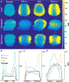
Figure 3.. Phantom SNR.
SNR data with 1 cm3 spatial resolution derived from 3-D echo-planar imaging of an ethylene glycol head phantom using: the birdcage transceiver; the 8-channel paddle coils separated by 19 cm (8ch19cm) to match the 32-channel housing; the 32-channel array; and the 8-channel paddle coils placed directly on the phantom, separated by 16.3 cm (8ch16.3cm)(A). Axial (white dashed line), mid-coronal, and mid-sagittal planes are shown, along with corresponding right-left (B), anterior-posterior (C), and superior-inferior (D) SNR line profiles, referenced by red dashed lines on (A); each profile point represents the mean of 3 adjacent voxels.
Figure 4.
Figure 4.. In vivo hyperpolarized data.
Hyperpolarized 2-D multi-slice EPI data having 4.5 cm3 spatial resolution acquired from a patient with a brain tumor using the 8- and 32-channel hardware configurations (8-channel paddles placed directly against the 15 cm head). Temporally-summed, parameter-normalized SNR>5 data for [1-13C]pyruvate (A) and [1-13C]lactate (B) are shown overlaid on 1H T1 post-contrast images. Ratios of mean slice SNR, [SNRmean,8-channel/SNRmean,32-channel]pyruvate and [SNRmean,8-channel/SNRmean,32-channel]lactate, were: 0.51,0.58; 0.99,1.02; 1.59,1.66; and 1.83,2.00 (superior-to-inferior slices). SNR>5 bicarbonate data depicts contrasting hardware sensitivity in the center of the brain (C).

References

    1. Ardenkjaer-Larsen JH, Fridlund B, Gram A, Hansson G, Hansson L, Lerche MH, Servin R, Thaning M, Golman K. Increase in signal-to-noise ratio of > 10,000 times in liquid-state NMR. Proc Natl Acad Sci USA. 2003; 100(18):10158–63 - PMC - PubMed
    1. Nelson SJ, Kurhanewicz J, Vigneron DB, Larson PEZ, Harzstark AL, Ferrone M, van Criekinge M, Chang JW, Bok R, Park I, Reed G, Carvajal L, Small EJ, Munster P, Weinberg VK, Ardenkjaer-Larsen JH, Chen AP, Hurd RE, Odegardstuen LI, Robb FJ, Tropp J, Murray JA. Metabolic imaging of patients with prostate cancer using hyperpolarized [1–13C]pyruvate. Sci Transl Med. 2013; 5(198): 198ra108 - PMC - PubMed
    1. Cunningham CH, Lau JYC, Chen AP, Geraghty BJ, Perks WJ, Roifman I, Wright GA, and Connelly KA. Hyperpolarized 13C Metabolic MRI of the Human Heart: initial experience. Circ Res. 2016; 119(11): 1177–1182 - PMC - PubMed
    1. Grist JT, McLean MA, Riemer F, Schulte RF, Deen SS, Zaccagna F, et al. Quantifying normal human brain metabolism using hyperpolarized [1–13C]pyruvate and magnetic resonance imaging. NeuroImage 2019; 189: 171–179 - PMC - PubMed
    1. Park I, Larson PEZ, Gordon JW, Carvajal L, Chen HY, Bok R, Van Criekinge M, Ferrone M, Slater JB, Xu D, Kurhanewicz J, Vigneron DB, Chang S, Nelson SJ. Development of methods and feasibility of using hyperpolarized carbon-13 imaging data for evaluating brain metabolism in patient studies. Magn Reson Med. 2018; 80(3):864–873 - PMC - PubMed

Publication types

MeSH terms