. 2019 Jun;10(3):696-709.
doi: 10.1002/jcsm.12412. Epub 2019 Mar 29.

4E-BP1 and 4E-BP2 double knockout mice are protected from aging-associated sarcopenia

Affiliations

4E-BP1 and 4E-BP2 double knockout mice are protected from aging-associated sarcopenia

Olivier Le Bacquer et al. J Cachexia Sarcopenia Muscle. 2019 Jun.

Abstract

Background: Sarcopenia is the loss of muscle mass/function that occurs during the aging process. The links between mechanistic target of rapamycin (mTOR) activity and muscle development are largely documented, but the role of its downstream targets in the development of sarcopenia is poorly understood. Eukaryotic initiation factor 4E-binding proteins (4E-BPs) are targets of mTOR that repress mRNA translation initiation and are involved in the control of several physiological processes. However, their role in skeletal muscle is still poorly understood. The goal of this study was to assess how loss of 4E-BP1 and 4E-BP2 expression impacts skeletal muscle function and homeostasis in aged mice and to characterize the associated metabolic changes by metabolomic and lipidomic profiling.

Methods: Twenty-four-month-old wild-type and whole body 4E-BP1/4E-BP2 double knockout (DKO) mice were used to measure muscle mass and function. Protein homeostasis was measured ex vivo in extensor digitorum longus by incorporation of l-[U-14 C]phenylalanine, and metabolomic and lipidomic profiling of skeletal muscle was performed by Metabolon, Inc.

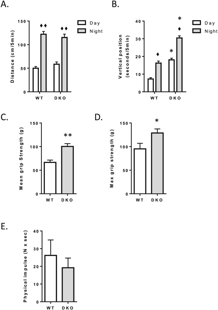
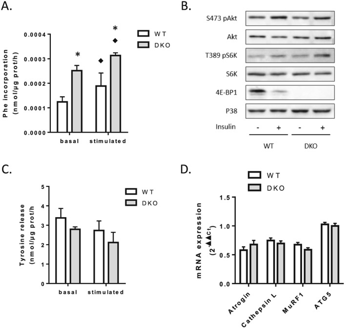
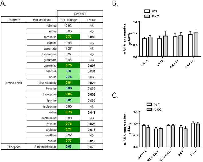
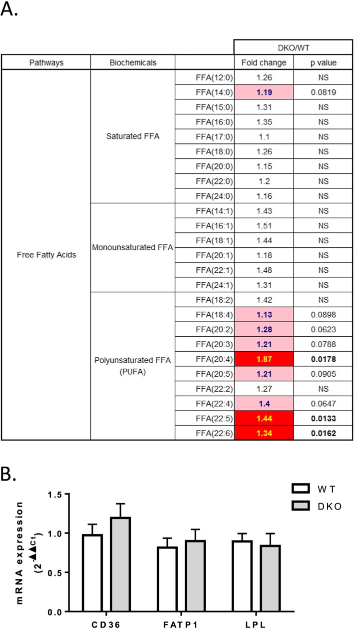
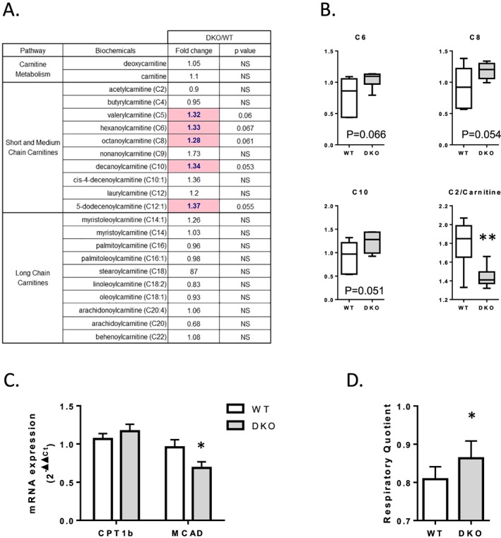
Results: The 4E-BP1/2 DKO mice exhibited an increase in muscle mass that was associated with increased grip strength (P < 0.05). Protein synthesis was higher under both basal (+102%, P < 0.05) and stimulated conditions (+65%, P < 0.05) in DKO skeletal muscle. Metabolomic and complex lipid analysis of skeletal muscle revealed robust differences pertaining to amino acid homeostasis, carbohydrate abundance, and certain aspects of lipid metabolism. In particular, levels of most free amino acids were lower within the 4E-BP1/2 DKO muscle. Interestingly, although glucose levels were unchanged, differences were observed in the isobaric compound maltitol/lactitol (33-fold increase, P < 0.01) and in several additional carbohydrate compounds. 4E-BP1/2 depletion also resulted in accumulation of medium-chain acylcarnitines and a 20% lower C2/C0 acylcarnitine ratio (P < 0.01) indicative of reduced β-oxidation.

Conclusions: Taken together, these findings demonstrate that deletion of 4E-BPs is associated with perturbed energy metabolism in skeletal muscle and could have beneficial effects on skeletal muscle mass and function in aging mice. They also identify 4E-BPs as potential targets for the treatment of sarcopenia.

Keywords: Anabolism; Protein synthesis; Proteolysis; Skeletal muscle; mTOR.


Conflict of interest statement

The authors declare they have no conflict of interest.

Figures

Figure 1
Figure 1
Spontaneous locomotor activity and muscle function in WT and 4E‐BP1/2 DKO mice. Spontaneous locomotor activity in WT and 4E‐BP1/2 DKO was measured using infrared sensor pairs arranged in strips for horizontal (A) and vertical (B) activities. Front forearm mean (C) and maximal (D) grip strength in WT and 4E‐BP1/2 DKO mice. (E) Mean holding impulse in the four limb wire grid holding test in WT and 4E‐BP1/2 DKO mice. n = 5–6 in each genotype, p values were assessed by two‐way analysis of variance, and Bonferroni post‐tests were used to compare replicate means by row (in A and B) or by unpaired t‐test (in CE). ♦P < 0.05 vs. day, ♦♦P < 0.01 vs. day, * P < 0.05 vs. WT, ** P < 0.01 vs. WT.
Figure 2
Figure 2
Loss of 4E‐BP1 and 4E‐BP2 alters protein homeostasis in skeletal muscle. (A) Protein synthesis in WT and 4E‐BP1/2 DKO skeletal muscles. Protein synthesis was measured ex vivo by radioactive phenylalanine incorporation in extensor digitorum longus with (stimulated) or without (basal) stimulation with a mixture of leucine and insulin. (B) Representative western blot levels of Akt, S6K, and 4E‐BP1 phosphorylation in WT and 4E‐BP1/2 DKO quadriceps. Mice were fasted overnight before receiving a 1.2 mU/g body weight intraperitoneal insulin injection. Mice were sacrificed 20 min later, and tissues were collected for western blot analysis. (C) Proteolysis in WT and 4E‐BP1/2 DKO skeletal muscles. Proteolysis was analysed ex vivo in extensor digitorum longus by measuring tyrosine release as described in the materials and methods Materials and methods. (D) Real‐time PCR quantification of Atrogin/MAFbx, Cathepsin L, MuRF1, and ATG5 mRNA expression in quadriceps from WT and 4E‐BP1/2 DKO mice. n = 5–6 in each genotype, p values were assessed by two‐way analysis of variance, and Bonferroni post‐tests were used to compare replicate means by row (in AC) or by unpaired t‐test (in D). ♦P < 0.05 vs. basal, * P < 0.05 vs. WT.
Figure 3
Figure 3
Free amino acid content is reduced in 4E‐BP1/2 DKO skeletal muscle. (A) Statistical heat map displaying the fold‐change values observed when amino acid abundances were compared between the WT and 4E‐BP1/2 DKO quadriceps of 24‐month‐old mice. The dark green colour is used to indicate statistically significant decreases P < 0.05, while the light green indicates differences that trended towards significance with a 0.05 < P < 0.1. (B) Real‐time PCR quantification of amino acid transporter mRNA expression in quadriceps from WT and 4E‐BP1/2 DKO 24‐month‐old mice. (C) Real‐time PCR quantification of mRNA expression of genes involved in branched‐chain amino acid catabolism in quadriceps from WT and 4E‐BP1/2 DKO 24‐month‐old mice. n = 6 in each genotype.
Figure 4
Figure 4
Loss of 4E‐BP1 and 4E‐BP2 induces accumulation of PUFA in skeletal muscle. (A) Statistical heat map displaying the fold‐change values observed when saturated, monounsaturated, and polyunsaturated free fatty acid abundance was compared between the quadriceps of 24‐month‐old WT and 4E‐BP1/2 DKO mice. The dark red colour is used to indicate statistically significant increases P < 0.05, while the light pink indicates differences that trended towards significance with a 0.05 < P < 0.1. (B) Real‐time PCR quantification of fatty acid transporters mRNA expression in quadriceps from WT and 4E‐BP1/2 DKO 24‐month‐old mice. n = 6 in each genotype.
Figure 5
Figure 5
Loss of 4E‐BP1 and 4E‐BP2 alters β‐oxidation in skeletal muscle. (A) Statistical heat map displaying the fold‐change values observed when short‐chain, medium‐chain, and long‐chain acylcarnitine abundance was compared between the quadriceps of 24‐month‐old WT and 4E‐BP1/2 DKO mice. The light pink indicates increases that trended towards significance with a 0.05 < P < 0.1. (B) Boxplots are shown for medium‐chain acylcarnitines and for the C2/carnitine ratio. (C) Real‐time PCR quantification of CPT1b and MCAD mRNA expression in quadriceps from WT and 4E‐BP1/2 DKO 24‐month‐old mice. (D) Respiratory quotient calculated as the ratio of VO2 to carbon dioxide production in WT and 4E‐BP1/2 DKO 24‐month‐old mice. *P < 0.05 vs. WT, **P < 0.01 vs. WT. n = 6 in each genotype.

References

    1. Saxton RA, Sabatini DM. mTOR signaling in growth, metabolism, and disease. Cell 2017;168:960–976. - PMC - PubMed
    1. Pause A, Belsham GJ, Gingras AC, Donze O, Lin TA, Lawrence JC Jr, et al. Insulin‐dependent stimulation of protein synthesis by phosphorylation of a regulator of 5′‐cap function. Nature 1994;371:762–767. - PubMed
    1. Poulin F, Gingras AC, Olsen H, Chevalier S, Sonenberg N. 4E‐BP3, a new member of the eukaryotic initiation factor 4E‐binding protein family. J Biol Chem 1998;273:14002–14007. - PubMed
    1. Haghighat A, Mader S, Pause A, Sonenberg N. Repression of cap‐dependent translation by 4E‐binding protein 1: competition with p220 for binding to eukaryotic initiation factor‐4E. EMBO J 1995;14:5701–5709. - PMC - PubMed
    1. Gingras AC, Gygi SP, Raught B, Polakiewicz RD, Abraham RT, Hoekstra MF, et al. Regulation of 4E‐BP1 phosphorylation: a novel two‐step mechanism. Genes Dev 1999;13:1422–1437. - PMC - PubMed

Publication types

MeSH terms