Review
. 2019 Mar;7(2):10.1128/microbiolspec.gpp3-0061-2019.
doi: 10.1128/microbiolspec.GPP3-0061-2019.

Immune Evasion by Staphylococcus aureus

Affiliations
Review

Immune Evasion by Staphylococcus aureus

Nienke W M de Jong et al. Microbiol Spectr. 2019 Mar.

Abstract

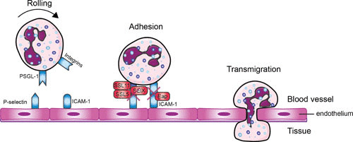
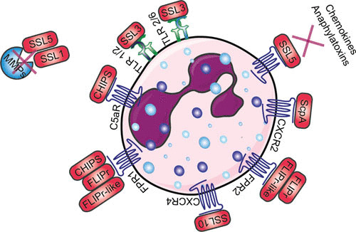
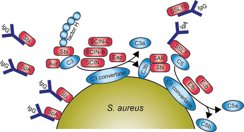
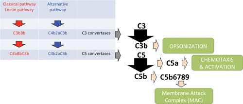
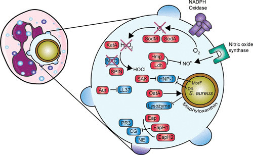
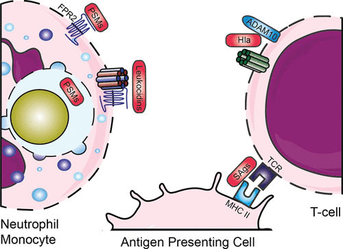
Staphylococcus aureus has become a serious threat to human health. In addition to having increased antibiotic resistance, the bacterium is a master at adapting to its host by evading almost every facet of the immune system, the so-called immune evasion proteins. Many of these immune evasion proteins target neutrophils, the most important immune cells in clearing S. aureus infections. The neutrophil attacks pathogens via a plethora of strategies. Therefore, it is no surprise that S. aureus has evolved numerous immune evasion strategies at almost every level imaginable. In this review we discuss step by step the aspects of neutrophil-mediated killing of S. aureus, such as neutrophil activation, migration to the site of infection, bacterial opsonization, phagocytosis, and subsequent neutrophil-mediated killing. After each section we discuss how S. aureus evasion molecules are able to resist the neutrophil attack of these different steps. To date, around 40 immune evasion molecules of S. aureus are known, but its repertoire is still expanding due to the discovery of new evasion proteins and the addition of new functions to already identified evasion proteins. Interestingly, because the different parts of neutrophil attack are redundant, the evasion molecules display redundant functions as well. Knowing how and with which proteins S. aureus is evading the immune system is important in understanding the pathophysiology of this pathogen. This knowledge is crucial for the development of therapeutic approaches that aim to clear staphylococcal infections.


Figures

FIGURE 1
FIGURE 1
Evading neutrophil extravasation to the infection site. Mechanisms by which S. aureus evades the steps in neutrophil extravasation. Neutrophils start to roll on the activated endothelium, which leads to firm adhesion and subsequently to transmigration through the endothelium. Red boxes indicate staphylococcal proteins, and blue boxes indicate host proteins. Abbreviations: PSGL-1, P-selectin glycoprotein 1; SSL, staphylococcal superantigen-like protein; ICAM-1, intracellular adhesion molecule 1; Eap, extracellular adherence protein; SElX, staphylococcal enterotoxin-like X. The figure was adapted from Servier Medical Art.
FIGURE 2
FIGURE 2
Schematic overview of how S. aureus evades priming, chemotaxis, and activation of neutrophils. Red boxes indicate staphylococcal proteins, and proteins shown in blue indicate host proteins. Abbreviations: TLR, Toll-like receptor; CXCR, chemokine receptor; ScpA, staphopain A; SSL, staphylococcal superantigen-like protein; FPR, formyl peptide receptor; FLIPr, FPR2 inhibitory protein; C5aR, C5a receptor; CHIPS, chemotaxis inhibitory protein of Staphylococcus; MMP, matrix metalloproteinase. The figure was adapted from Servier Medical Art.
FIGURE 3
FIGURE 3
Diagram of the main pathways and components of the human complement system 3a and a schematic representation of S. aureus evading opsonization and phagocytic uptake by neutrophil 3b. Red boxes indicate staphylococcal proteins, and blue boxes indicate host proteins. Abbreviations: IgG, immunoglobulin G; SpA, staphylococcal protein A; Sbi, staphylococcal binding of IgG; SCIN, staphylococcal complement inhibitor; SAK, staphylokinase; Aur, aureolysin; SSL, staphylococcal superantigen-like protein; Efb, extracellular fibrinogen-binding protein; Ecb, extracellular complement-binding protein. The figure was adapted from Servier Medical Art.
FIGURE 3
FIGURE 3
Diagram of the main pathways and components of the human complement system 3a and a schematic representation of S. aureus evading opsonization and phagocytic uptake by neutrophil 3b. Red boxes indicate staphylococcal proteins, and blue boxes indicate host proteins. Abbreviations: IgG, immunoglobulin G; SpA, staphylococcal protein A; Sbi, staphylococcal binding of IgG; SCIN, staphylococcal complement inhibitor; SAK, staphylokinase; Aur, aureolysin; SSL, staphylococcal superantigen-like protein; Efb, extracellular fibrinogen-binding protein; Ecb, extracellular complement-binding protein. The figure was adapted from Servier Medical Art.
FIGURE 4
FIGURE 4
Overview of evasion proteins that are involved in evading neutrophil killing. Enlargement of the phagosome is shown on the right. Red boxes indicate staphylococcal proteins, and blue boxes indicate host proteins. Staphyloxanthin provides a protective shield, KatA neutralizes hydrogen peroxide (H2O2) into water (H2O) and oxygen (O2), and SPIN inhibits MPO activity. MprF and the Dlt operon lead to an increase in positive charge of the bacterial surface. Abbreviations: SOD, superoxide dismutase; SAK, staphylokinase; KatA, catalase; MPO, myeloperoxidase; SPIN, staphylococcal peroxidase inhibitor; Aur, aureolysin; Hmp, flavohemoglobin; Ldh, l-lactate dehydrogenase; Eap, extracellular adherence protein; EapH, extracellular adherence protein homologue; PR3, proteinase 3; CG, cathepsin G; NE, neutrophil elastase. The figure was adapted from Servier Medical Art.
FIGURE 5
FIGURE 5
Evasion by staphylococcal toxins. Various leukocidins bind specific GPCRs, after which they form a pore and lyse host cells. PSMs are released inside the phagosome and can bind via FPR2. SAgs cross-link major histocompatibility complex class II and T-cell receptors. Abbreviations: GPCR, G-protein-coupled receptor; FPR, formyl protein receptor; PSMs, phenol-soluble modulins; Hla, hemolysin-alpha; SAgs, superantigens; MHC II, major histocompatibility complex II; TCR, T-cell receptor. The figure was adapted from Servier Medical Art.

References

    1. Gorwitz RJ, Kruszon-Moran D, McAllister SK, McQuillan G, McDougal LK, Fosheim GE, Jensen BJ, Killgore G, Tenover FC, Kuehnert MJ. 2008. Changes in the prevalence of nasal colonization with Staphylococcus aureus in the United States, 2001-2004. J Infect Dis 197:1226–1234 10.1086/533494. [PubMed] - DOI - PubMed
    1. Krismer B, Weidenmaier C, Zipperer A, Peschel A. 2017. The commensal lifestyle of Staphylococcus aureus and its interactions with the nasal microbiota. Nat Rev Microbiol 15:675–687 10.1038/nrmicro.2017.104. [PubMed] - DOI - PubMed
    1. von Eiff C, Becker K, Machka K, Stammer H, Peters G. 2001. Nasal carriage as a source of Staphylococcus aureus bacteremia. N Engl J Med 344:11–16 10.1056/NEJM200101043440102. [PubMed] - DOI - PubMed
    1. Lowy FD. 1998. Staphylococcus aureus infections. N Engl J Med 339:520–532 10.1056/NEJM199808203390806. [PubMed] - DOI - PubMed
    1. Barrett FF, McGehee RF Jr, Finland M. 1968. Methicillin-resistant Staphylococcus aureus at Boston City Hospital. Bacteriologic and epidemiologic observations. N Engl J Med 279:441–448 10.1056/NEJM196808292790901. [PubMed] - DOI - PubMed

Publication types

MeSH terms

LinkOut - more resources