. 2019 Mar 30;18(1):68.
doi: 10.1186/s12943-019-0972-8.

Targeting interleukin-6 as a strategy to overcome stroma-induced resistance to chemotherapy in gastric cancer

Affiliations

Targeting interleukin-6 as a strategy to overcome stroma-induced resistance to chemotherapy in gastric cancer

In-Hye Ham et al. Mol Cancer. .

Abstract

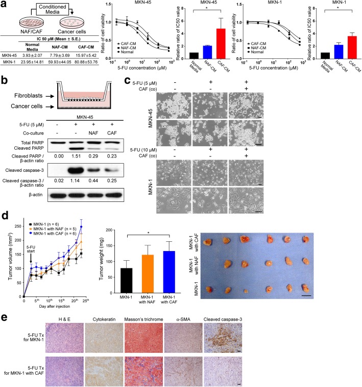
Background: Although the tumor stroma in solid tumors like gastric cancer (GC) plays a crucial role in chemo-resistance, specific targets to inhibit the interaction between the stromal and cancer cells have not yet been utilized in clinical practice. The present study aims to determine whether cancer-associated fibroblasts (CAFs), a major component of the tumor stroma, confer chemotherapeutic resistance to GC cells, and to discover potential targets to improve chemo-response in GC.

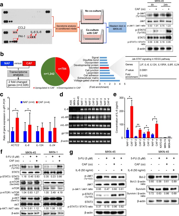
Methods: To identify CAF-specific proteins and signal transduction pathways affecting chemo-resistance in GC cells, secretome and transcriptome analyses were performed. We evaluated the inhibiting effect of CAF-specific protein in in vivo and in vitro models and investigated the expression of CAF-specific protein in human GC tissues.

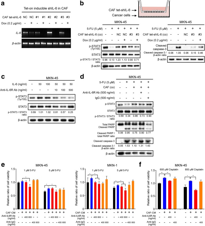
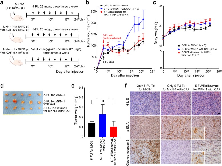
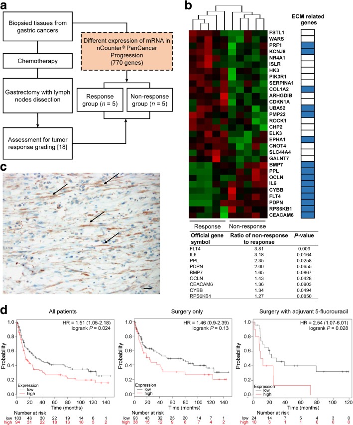
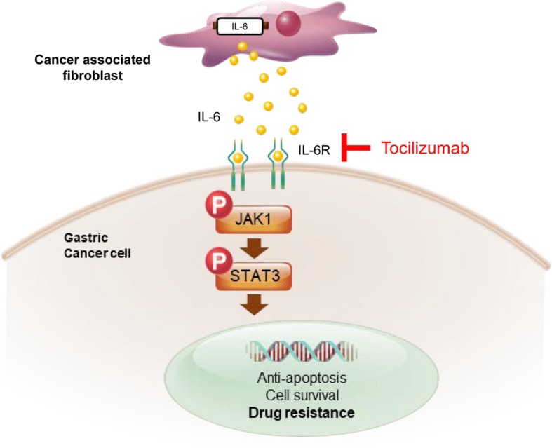
Results: Secretome and transcriptome data revealed that interleukin-6 (IL-6) is a CAF-specific secretory protein that protects GC cells via paracrine signaling. Furthermore, CAF-induced activation of the Janus kinase 1-signal transducer and activator of transcription 3 signal transduction pathway confers chemo-resistance in GC cells. CAF-mediated inhibition of chemotherapy-induced apoptosis was abrogated by the anti-IL-6 receptor monoclonal antibody tocilizumab in various experimental models. Clinical data revealed that IL-6 was prominently expressed in the stromal portion of GC tissues, and IL-6 upregulation in GC tissues was correlated with poor responsiveness to chemotherapy.

Conclusions: Our data provide plausible evidence for crosstalk between GC cells and CAFs, wherein IL-6 is a key contributor to chemoresistance. These findings suggest the potential therapeutic application of IL-6 inhibitors to enhance the responsiveness to chemotherapy in GC.

Keywords: Cancer-associated fibroblasts; Chemo-resistance; Gastric cancer; Interleukin-6; Jak1-STAT3; Tocilizumab; Tumor microenvironment.


Conflict of interest statement

Ethics approval and consent to participate

This study was approved by the institutional review board/ethics committee of the Ajou University Hospital (AJIRB-BMR-KSP-15-432).

Consent for publication

Not applicable.

Competing interests

R.A. Brekken has an ownership interest (including stock, patents, etc.) in and is a consultant/advisory board member for Tuevol Therapeutics. Other authors declare that they have no competing interests.

Publisher’s Note

Springer Nature remains neutral with regard to jurisdictional claims in published maps and institutional affiliations.

Figures

Fig. 1
Fig. 1
Cancer-associated fibroblast (CAF)-induced resistance to 5-fluorouracil (5-FU) in gastric cancer cells. a MKN-45 and MKN-1 gastric cancer cells treated with 5-FU were treated with fibroblast culture-conditioned media, and the half maximal inhibitory concentration (IC50) was measured. The results are presented as the mean (± SEM). *P < 0.05, based on Kruskal-Wallis test followed by a Dunn’s multiple comparison. b Schematic figure detailing the transwell co-culture system with fibroblasts isolated from paired normal gastric tissues (normal-associated fibroblasts or NAFs) and gastric cancer tissues (CAFs). Western blot analysis results show changes in the expression of apoptotic markers such as cleaved PARP and caspase-3 72 h after 5-FU treatment with and without co-culture with NAFs and CAFs. c Representative micrographs demonstrating morphological changes in MKN-45 and MKN-1 cells after 5-FU treatment for 72 h with and without co-culture with fibroblasts. Scale bar = 100 μm. d A line graph comparing tumor growth among the in vivo xenograft tumors derived from MKN-1 cells alone (n = 6), MKN-1 cells combined with NAFs (n = 5), or MNK-1 cells combined with CAFs (n = 5) after 5-FU treatments. The bar graph compares the harvested tumor weight among the three groups. Graphs show the mean (± SEM) tumor weights of the mice. *P < 0.05, based on one-way ANOVA analysis, followed by a post hoc test with Tukey’s method. The photographs show the harvested tumors. Scale bar = 1 cm. e. Representative micrographs showing H&E staining, Masson’s trichrome staining for stromal collagen fibers, and immunohistochemical staining for α-smooth muscle actin (a-SMA), cytokeratin, and cleaved caspase-3 in harvested xenograft tumors derived from only MKN-1 cells and those derived from MKN-1 cells mixed with CAFs after treatment with 5-FU. Scale bar = 100 μm
Fig. 2
Fig. 2
Identification of the IL-6/Jak1/STAT3 axis as a specific communicator between cancer-associated fibroblasts (CAFs) and gastric cancer cells. a Interleukin-6 (IL-6), interleukin-8 (IL-8), and C-C motif chemokine ligand 2 (CCL2) were secreted at higher levels in the media after co-culture with CAFs and MKN-45 cells than in the media used for culturing MKN-45 cells alone. Each of these factors were correlated with the Jak-STAT3 signal transduction pathway. The Western blot analysis shows expression changes of the indicated proteins with and without co-culture with CAFs for 6 or 24 h. b A flow chart depicting the transcriptome analysis of one paired set of normal-associated fibroblasts (NAFs) and CAFs. The pie graph presents the number of upregulated and downregulated genes in CAFs compared with those in NAFs. The graph and table show the functional annotation of results for 784 upregulated genes in CAFs from the Database for Annotation, Visualization, and Integrated Discovery (DAVID: https://david.ncifcrf.gov), which is based on the Kyoto Encyclopedia of Genes and Genomes pathways. c Results from quantitative PCR (qPCR) analysis showing the comparative mRNA expression of ACTC2, IL6, IL12A, and IL24 between the NAFs and CAFs. The graphs show the mean (± SEM) ratio of mRNA expression in CAFs compared to those in NAFs. *P < 0.05, based on paired t-tests. d Reverse transcription PCR (RT-PCR) results showing mRNA expression of IL-6 and its receptors in cells from the gastric cancer cell lines AGS, MKN-1, and MKN-45, and four paired NAFs and CAFs. e ELISA results showing IL-6 levels in the conditioned media from four paired NAFs and CAFs and from cells of the gastric cancer cell lines KATO-III, MKN-28, and MKN-45. *P < 0.05 and **P < 0.001, according to paired t-tests. f, g Western blot analysis showing the expression levels of the indicated proteins after 5-fluorouracil (5-FU) treatment (5 μM) with and without co-culture with CAFs and with and without recombinant IL-6 treatment in MKN-45 cells and MKN-1 cells
Fig. 3
Fig. 3
Suppressive effect of interleukin-6 (IL-6) inhibition on the cancer-associated fibroblast (CAF)-induced resistance to 5-fluorouracil (5-FU). a Reverse transcription (RT)-PCR analysis showing the expression of IL6 and ACTB mRNA in CAFs transfected with three different tet-on inducible IL6 shRNAs vectors or a negative control vector [38]. Dox indicates doxycycline. b Schematic figure depicting the transwell co-culture system for tet-on IL6 shRNA-transfected CAFs and gastric cancer cells. Western blot analysis shows the expression of the apoptotic markers cleaved PARP, caspase-3, and phosphorylated STAT3 in the lysate of MKN-45 cell cultures in the lower chamber after doxycycline (0.2 μg/ml) treatment of CAFs transfected with the tet-on IL6 shRNA or negative control (NC) vector in the upper chamber. c Western blot analysis showing the expression of the indicated proteins in cells treated with human recombinant IL-6 combined with and without tocilizumab treatment. d Western blot analysis showing the expression of the indicated proteins in the lysates from MKN-45 and MKN-1 cells after 5-FU (5 μM) treatment with and without CAFs and subsequent treatment with tocilizumab (500 ng/ml) or negative control IgG (500 ng/ml). e Ez-cytox tests showing the relative ratio of the viability of MKN-45 and MKN-1 cells treated with 1 μM or 5 μM of 5-FU after the addition of tocilizumab (400 and 800 ng/ml) or control IgG (400 and 800 ng/ml). f Ez-cytox tests demonstrating the relative ratio of cell viability in MKN-45 cultures treated with 600 μM or 800 μM cisplatin after the addition of tocilizumab (400 ng/ml) or control IgG (400 ng/ml). The graphs show the mean (± SEM) ratios of cell viability. *P < 0.05 and **P < 0.001, according to Mann-Whitney test
Fig. 4
Fig. 4
Effect of anti-interleukin-6 (IL-6) receptor monoclonal antibody on 5-fluorouracil (5-FU) treatment of mixed xenograft tumors derived from cancer-associated fibroblasts (CAFs). a The panels show representative images of the samples from each group. The arrows indicate the days on which the treatment was administered. b A line graph displaying the comparison of tumor growth among the in vivo xenograft tumors derived from MKN-1 cells alone (n = 5) and MKN-1 cells combined with CAFs (n = 5) after 5-FU treatments, and xenograft tumors derived from MKN-1 cells combined with CAFs (n = 5) treated simultaneously with 5-FU and tocilizumab. c A line graph showing the changes in body weight among the three groups of mice. d The photographs show the tumor-bearing mice before euthanasia and the harvested tumors. e The column graph comparing the harvested tumor weights among the three groups. The graphs show the mean (± SEM) tumor weights of the mice. *P < 0.05 and **P < 0.001, according to Mann-Whitney test. f Representative micrographs of the tumors harvested from the mice in the three groups; the tumor samples were analyzed by H&E staining and immunohistochemical staining for α-SMA and cleaved caspase-3. Scale bar = 100 μm
Fig. 5
Fig. 5
Gene expression pattern in pre-treatment biopsied tissues of patients who underwent preoperative chemotherapy. a Flow diagram presenting the study scheme for the comparison of gene expression patterns in the pre-treatment biopsied gastric cancer tissues between patients in the chemotherapy response and non-response groups. b Heatmap showing the top nine genes, including interleukin-6 (IL-6), usually those associated with the extracellular matrix layer, and the expression pattern of these genes appropriately clustered into the response and non-response patient groups. A list of upregulated genes is shown for the non-response group when gene expression was evaluated in biopsied tissues from primary tumors of pretreated gastric cancer patients c Representative micrograph of IL-6 immunohistochemical staining of gastric cancer tissues showing IL-6 expression specifically in the stromal cells (black arrows), but not in the cancer cells. Scale bar = 100 μm. d Disease-free survival assessed using the GSE15459 gastric cancer dataset at www.kmplot.com. The difference in survival relative to IL6 mRNA expression was compared in each group involving all patients, i.e., those treated with surgery only and those treated with surgery and adjuvant chemotherapy, according to Kaplan-Meier survival analysis with log-rank test
Fig. 6
Fig. 6
Schematic figure for present study. Cancer-associated fibroblast (CAF)-induced interleukin-6 (IL-6) activates the Jak1-STAT3 pathway in gastric cancer cells via paracrine signaling, which allows tumor cells to increasingly oppose apoptosis and increase their survival and resistance to chemotherapy. Tocilizumab, a humanized monoclonal anti-IL-6R antibody that is an FDA-approved drug, inhibits the CAF-induced activation of the Jak1-STAT3 signaling pathway in gastric cancer cells and consequently increases the efficacy of chemotherapeutic drugs

References

    1. Ferlay J, Soerjomataram I, Dikshit R, Eser S, Mathers C, Rebelo M, Parkin DM, Forman D, Bray F. Cancer incidence and mortality worldwide: sources, methods and major patterns in GLOBOCAN 2012. Int J Cancer. 2015;136:E359–E386. doi: 10.1002/ijc.29210. - DOI - PubMed
    1. Glimelius B, Ekstrom K, Hoffman K, Graf W, Sjoden PO, Haglund U, Svensson C, Enander LK, Linne T, Sellstrom H, Heuman R. Randomized comparison between chemotherapy plus best supportive care with best supportive care in advanced gastric cancer. Ann Oncol. 1997;8:163–168. doi: 10.1023/A:1008243606668. - DOI - PubMed
    1. Murad AM, Santiago FF, Petroianu A, Rocha PR, Rodrigues MA, Rausch M. Modified therapy with 5-fluorouracil, doxorubicin, and methotrexate in advanced gastric cancer. Cancer. 1993;72:37–41. doi: 10.1002/1097-0142(19930701)72:1<37::AID-CNCR2820720109>3.0.CO;2-P. - DOI - PubMed
    1. Pyrhonen S, Kuitunen T, Nyandoto P, Kouri M. Randomised comparison of fluorouracil, epidoxorubicin and methotrexate (FEMTX) plus supportive care with supportive care alone in patients with non-resectable gastric cancer. Br J Cancer. 1995;71:587–591. doi: 10.1038/bjc.1995.114. - DOI - PMC - PubMed
    1. Housman G, Byler S, Heerboth S, Lapinska K, Longacre M, Snyder N, Sarkar S. Drug resistance in cancer: an overview. Cancers (Basel) 2014;6:1769–1792. doi: 10.3390/cancers6031769. - DOI - PMC - PubMed

Publication types

MeSH terms