Review
. 2019 Mar 30;18(1):66.
doi: 10.1186/s12943-019-0962-x.

Stem cells in homeostasis and cancer of the gut

Affiliations
Review

Stem cells in homeostasis and cancer of the gut

Maartje van der Heijden et al. Mol Cancer. .

Abstract

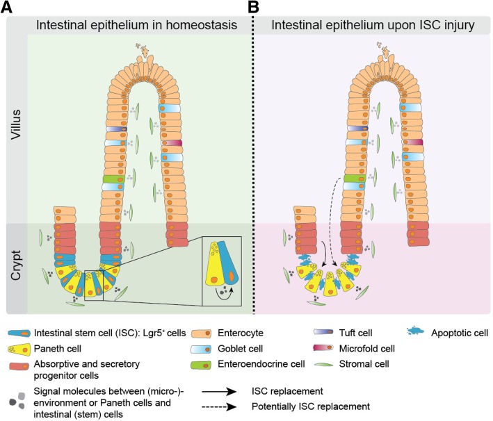
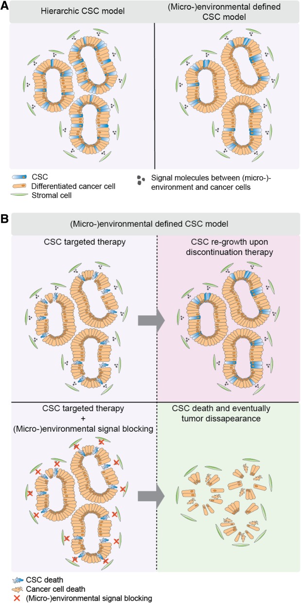
The intestinal epithelial lining is one of the most rapidly renewing cell populations in the body. As a result, the gut has been an attractive model to resolve key mechanisms in epithelial homeostasis. In particular the role of intestinal stem cells (ISCs) in the renewal process has been intensely studied. Interestingly, as opposed to the traditional stem cell theory, the ISC is not a static population but displays significant plasticity and in situations of tissue regeneration more differentiated cells can revert back to a stem cell state upon exposure to extracellular signals. Importantly, normal intestinal homeostasis provides important insight into mechanisms that drive colorectal cancer (CRC) development and growth. Specifically, the dynamics of cancer stem cells bear important resemblance to ISC functionality. In this review we present an overview of the current knowledge on ISCs in homeostasis and their role in malignant transformation. Also, we discuss the existence of stem cells in intestinal adenomas and CRC and how these cells contribute to (pre-)malignant growth. Furthermore, we will focus on new paradigms in the field of dynamical cellular hierarchies in CRC and the intimate relationship between tumor cells and their niche.

Keywords: Cancer stem cells; Cell plasticity; Colorectal cancer; Intestinal stem cells; Tumor (micro-)environment.


Conflict of interest statement

Ethics approval and consent to participate

Not applicable.

Consent for publication

Not applicable.

Competing interests

The authors declare that they have no competing interests.

Publisher’s Note

Springer Nature remains neutral with regard to jurisdictional claims in published maps and institutional affiliations.

Figures

Fig. 1
Fig. 1
The intestinal epithelium. (a) The intestinal lining consists of an epithelial monolayer covering invaginations (crypts) and finger like protrusions (villi, only in the small intestine). Intestinal stem cells (ISC) reside in the bottom of the crypts, absorptive and secretory progenitor cells directly above the ISC zone, and more differentiated cells towards the top of crypts and on the villi. Intestinal progenitor and differentiated cells move upwards due to the massive tissue renewal fueled by the ISCs. This is a continuous process and it only takes several days before differentiated cells undergo apoptosis and are shed into the gut lumen. (b) The ISC compartment is sensitive to cytotoxic injury, such as irradiation. Consequently, upon DNA damage ISCs undergo apoptosis. The progenitor cells located higher up in the crypt replace the loss of ISCs and due to the new topological position regain niche signals, which then install ISC activity. Therefore, the ISC compartment is a dynamic population and progenitor- and potentially fully differentiated cells, show an enormous cellular plasticity upon ISC loss
Fig. 2
Fig. 2
Intestinal cell plasticity dynamics in malignant transformation. (a) ISCs have the ability to effectively initiate adenoma formation when these cells acquire Apc mutations. On the other hand, differentiated intestinal epithelial cells do rarely undergo malignant transformation upon obtaining an oncogenic hit. (b) However, in an inflammatory environment differentiated cells acquire similar transformation potential. Different factors underlie the differences in transformation capacity of ISCs versus differentiated cells. First of all, the topological position of differentiated cells prevents them in homeostasis from generating long-lived clonal lineages. Secondly, the ISC niche endows ISCs with the potential to endure the stressors that result from acquiring an oncogenic mutation [74]. Similarly, in a colitis environment the differentiated cell compartment is also installed with anti-apoptotic capacities through activation of the nuclear factor-κB (Nf-κB) pathway [73]. The anti-apoptotic protein BLC-2 is one of the identified mediators that facilitates this oncogenic transformation. Indeed, inhibition of BCL-2, either genetically or pharmacologically, reduced adenoma burden in mice [74]
Fig. 3
Fig. 3
Cancer stem cell dynamics in colorectal cancer growth and therapy. (a) The strictly hierarchical cancer stem cell (CSC) model postulates that the CSC state is a fixed entity and CSCs are intrinsically equipped with self-renewal potential and multi-potency. On the opposite, the (micro-)environmental defined CSC model states that signal molecules derived from the stromal compartment install CRC cells with CSC potential, such as self-renewal and multi-lineage differentiation capacity. (b) The top panel predicts that eradicating CSCs by blocking important stem cell signaling pathways, e.g. Wnt signaling, is not sufficient to halt tumor growth. Once treatment is discontinued (top left panel) specific CSC-installing signals from the niche will provide CRC cells with CSC potential and these CSCs will again drive tumor growth. The bottom panel shows a situation in which both (micro-)environmental signals as CSC-specific pathways are blocked resulting in potentially effective tumor control

References

    1. Siegel RL, Miller KD, Fedewa SA, Ahnen DJ, Meester RGS, Barzi A, et al. Colorectal cancer statistics, 2017. CA Cancer J Clin. 2017;67(3):177–193. doi: 10.3322/caac.21395. - DOI - PubMed
    1. Hanahan D, Weinberg RA. The hallmarks of cancer. Cell. 2000;100(1):57–70. doi: 10.1016/S0092-8674(00)81683-9. - DOI - PubMed
    1. Fearon ER, Vogelstein B. A genetic model for colorectal tumorigenesis. Cell. 1990;61(5):759–767. doi: 10.1016/0092-8674(90)90186-I. - DOI - PubMed
    1. de Sousa e Melo F, Kurtova AV, Harnoss JM, Kljavin N, Hoeck JD, Hung J, et al. A distinct role for Lgr5(+) stem cells in primary and metastatic colon cancer. Nature. 2017;543(7647):676–680. - PubMed
    1. Lenos KJ, Miedema DM, Lodestijn SC, Nijman LE, van den Bosch T, Romero Ros X, et al. Stem cell functionality is microenvironmentally defined during tumour expansion and therapy response in colon cancer. Nat Cell Biol. 2018;20(10):1193–1202. doi: 10.1038/s41556-018-0179-z. - DOI - PMC - PubMed

Publication types