Review
. 2019 May 15:666:40-45.
doi: 10.1016/j.abb.2019.03.013. Epub 2019 Mar 27.

Invertebrate troponin: Insights into the evolution and regulation of striated muscle contraction

Affiliations
Review

Invertebrate troponin: Insights into the evolution and regulation of striated muscle contraction

Tianxin Cao et al. Arch Biochem Biophys. .

Abstract

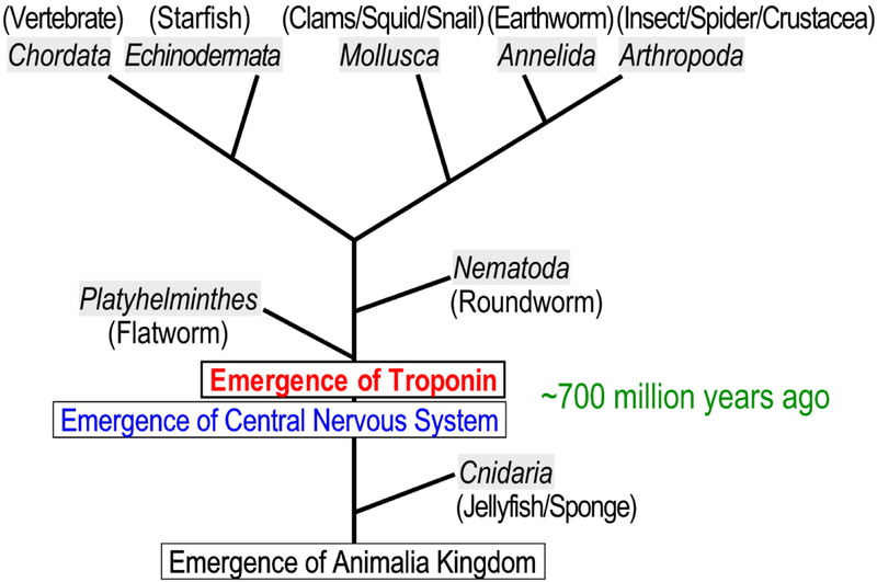
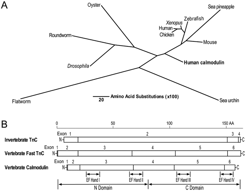
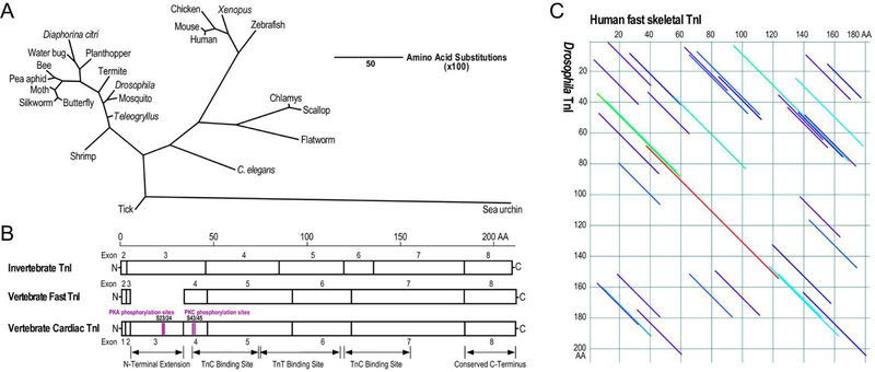
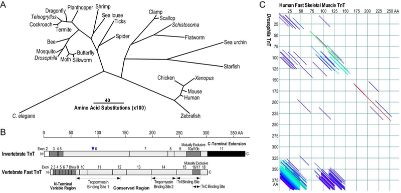
The troponin complex plays a central role in regulating the contraction and relaxation of striated muscles. Among the three protein subunits of troponin, the calcium receptor subunit, TnC, belongs to the calmodulin family of calcium signaling proteins whereas the inhibitory subunit, TnI, and tropomyosin-binding/thin filament-anchoring subunit, TnT, are striated muscle-specific regulatory proteins. TnI and TnT emerged early in bilateral symmetric invertebrate animals and have co-evolved during the 500-700 million years of muscle evolution. To understand the divergence as well as conservation of the structures of TnI and TnT in invertebrate and vertebrate organisms adds novel insights into the structure-function relationship of troponin and the muscle type isoforms of TnI and TnT. Based on the significant growth of genomic database of multiple species in the past decade, this focused review studied the primary structure features of invertebrate troponin subunits in comparisons with the vertebrate counterparts. The evolutionary data demonstrate valuable information for a better understanding of the thin filament regulation of striated muscle contractility in health and diseases.

Keywords: Invertebrate muscle; Molecular evolution; Myofilament; TnI; TnT; Troponin isoforms.


Figures

Fig. 1.
Fig. 1.. Early emergence of troponin during the evolution of animals.
Based on available sequence data of animal genomes, this unscaled illustration of phylogenic lineage of the phyla of animal kingdom illustrates the emergence of troponin ~700 million years ago prior to the emergence of Platyhelminthes and Nematoda.
Fig. 2.
Fig. 2.. Evolution and structure features of TnC in vertebrates and invertebrates.
(A) The phylogenetic tree of TnC constructed by aligning amino acid sequences from representative species of animal phyla using the Clustal V method of DNAStar MegAlign software shows the divergence of TnC between invertebrates and vertebrates as well as the homology to calmodulin. The length of each pair of branches represents the distance between sequence pairs, while the scale bar indicating the distance corresponding to 20 amino acid substitutions per 100 residues. The sequence accession numbers are: Sea urchin TnC, AAA30007.1; flatworm TnC, XP_012797142.1; Drosophila TnC, NP_523619.2; roundworm TnC, BAA82523.1; Oyster TnC, BBD82024.1; chicken TnC, AAA49097.1; human TnC, AAA91854.1; Xenopus TnC, NP_001079408.1; zebrafish TnC, AAH64284.1; sea pineapple TnC, BAA13630.1; mouse TnC, NP_033419; human calmodulin, CAA36839. (B) The linear maps of protein primary structures illustrate the similarity between invertebrate and vertebrate TnC and to calmodulin with the four EF hand metal binding sites in the N and C domains indicated.
Fig. 3.
Fig. 3.. Evolution and structure features of TnI in vertebrates and invertebrates.
(A) The phylogenetic tree of TnI constructed by aligning amino acid sequences from representative species of animal phyla using the Clustal V method of DNAStar MegAlign software demonstrates significant evolutionary divergence while vertebrates and insects being conserved clusters. The length of each pair of branches represents the distance between sequence pairs, while the scale bar indicating the distance corresponding to 50 amino acid substitutions per 100 residues. The sequence accession numbers are: Sea urchin TnI, XP_011664558.1; tick TnI, BAB55451.1; shrimp TnI, ACV40756.1; butterfly TnI, NP_001299300.1; silkworm TnI, NP_001037295.1; moth TnI, KOB71297.1; pea aphid TnI, NP_001313576.1; bee TnI, NP_001035346.1; water bug TnI, CAF18234.1; Diaphorina citri TnI, ABG81999.1; planthopper TnI, ACN79503.1; termite TnI, KDQ88314.1; Drosophila TnI, CAA42020.1; mosquito TnI, XP_001864736.1; teleogryllus TnI, AVI126882.1; human fast TnI, AAH32148.1; mouse TnI, NP_033431.1; chicken fast TnI, AAA61952.1; Xenopus fast TnI, AAH84508.1; zebrafish fast TnI, AAI62242.1; chlamys TnI, BAE43658.1; scallop TnI, BAE43658.1; flatworm TnI CAX73588.1; C. elegans TnI, NP_509906.1. B) The linear maps of protein primary structure demonstrate the conserved core structures of vertebrate and invertebrate TnI containing TnC and TnT binding sites. A regulatory N-terminal extension is present in vertebrate cardiac TnI. Invertebrate TnI also has an N-terminal extension although its function has not been extensively studied. C) Paired DotPlot alignment of amino acid sequences of human fast skeletal muscle TnI (accession # AAH32148.1) and Drosophila TnI (accession # CAA42020.1) demonstrates the conserved core structures. The diagonal lines indicate regions of the two sequences which meet the threshold for similarity specified in the parameters set for the analysis, of which dark blue indicates weak similarity with progressively stronger similarities shown in light blue, green, yellow, orange and red. The long red diagonal line indicates the region containing conserved binding sites for TnC and TnT (B).
Fig. 4.
Fig. 4.. Evolution and structure features of TnT in vertebrates and invertebrates.
(A) The phylogenetic tree of TnT constructed by aligning amino acid sequences from representative species in animal phyla using the Clustal V method of DNAStar MegAlign software shows significant evolutionary divergence. The length of each pair of branches represents the distance between sequence pairs, while the scale bar indicating the distance corresponding to 40 amino acid substitutions per 100 residues. Similar to the pattern of TnI shown in Fig. 3A, vertebrate TnT and insect TnT are conserved in clusters. The sequence accession numbers are: C. elegans TnT, NP_001024704.1; butterfly TnT, BAG30738.1; silkworm TnT, ABD36267.1; moth TnT, ADO33067.1; Drosophila TnT, NP_001162742.1; mosquito TnT, XP_001851541.1; bee TnT, NP_001035348.1; termite TnT, AGM32088.1; cockroaches TnT, AAD33603.1; Teleogryllus TnT, AVI26881.1; dragonfly TnT, AAD33604.1; planthopper TnT, AGI96988.1; shrimp TnT, AQV08184.2; sea louse TnT, ACO12887.1; tick TnT, AAY42205.1; spider TnT, EU247211.1; clamp TnT, BAA13610.1; scallop TnT, BAA20456.1; Schistosoma TnT, XP_018646291.1; flatworm TnT, XP_024354240.1; sea urchin TnT, XP_011671209.1; ; starfish TnT, XP_022088338.1; chicken TnT, NP_990253.1; Xenopus TnT, NP_989143.1; mouse TnT, NP_001347086.1; human TnT, NP_001354775.1; fish TnT, AAF78472.1. B) The linear maps of protein primary structure outline the divergence as well as conservation between vertebrate and invertebrate TnT with their middle and C-terminal conserved regions (highlighted in gray) containing binding sites for tropomyosin, TnI and TnC, an N-terminal variable region, a C-terminal mutually spliced segment, and a C-terminal extension unique to invertebrate TnT, especially insect TnT. The alternatively spliced exons are shown as hatched boxes whereas the C-terminal extension of invertebrate TnT is highlighted in solid black. The alternative translational initiation site that generates N-terminal truncated TnT in Drosophila is indicated with a blue arrow. C) Paired DotPlot alignment of amino acid sequences of human fast skeletal muscle TnT (accession # NP_001354775.1) and Drosophila TnT (accession # NP_001162742.1) demonstrates the middle and C-terminal conserved regions. The diagonal lines indicate regions of the two sequences which meet the threshold for similarity specified in the parameters set for the analysis, of which dark blue indicates weak similarity with progressively stronger similarities shown in light blue, green, yellow, orange and red. Although non-homologous structures, the N-terminal variable region of vertebrate TnT and the C-terminal extension of insect TnT showed a strong similarity based on their unique high contents of Glu residues.

References

    1. Sanger JW, Wang J, Fan Y, White J, Mi-Mi L, Dube DK, Sanger JM, Pruyne D. Assembly and maintenance of myofibrils in striated muscle. Handbook of Experimental Pharmacology. 2017; 235:39–75. - PubMed
    1. Perkins AD, Tanentzapf G. An ongoing role for structural sarcomeric components in maintaining Drosophila melanogaster muscle function and structure. PLoS One. 2014; 9:e99362. - PMC - PubMed
    1. Paniagua R, Royuela M, García-Anchuelo RM, Fraile B. Ultrastructure of invertebrate muscle cell types. Histol Histopathol. 1996; 11:181–201. - PubMed
    1. Tobacman LS. Thin filament-mediated regulation of cardiac contraction. Annu Rev Physiol. 1996; 58:447–481. - PubMed
    1. Gordon AM, Homsher E, Regnier M. Regulation of contraction in striated muscle. Physiol Rev. 2000; 80:853–924. - PubMed

Publication types

LinkOut - more resources