Clinical Trial
. 2019:22:101792.
doi: 10.1016/j.nicl.2019.101792. Epub 2019 Mar 26.

Cause or effect: Altered brain and network activity in cervical dystonia is partially normalized by botulinum toxin treatment

Affiliations
Clinical Trial

Cause or effect: Altered brain and network activity in cervical dystonia is partially normalized by botulinum toxin treatment

Stefan Brodoehl et al. Neuroimage Clin. 2019.

Abstract

Background: Idiopathic cervical dystonia (CD) is a chronic movement disorder characterized by impressive clinical symptoms and the lack of clear pathological findings in clinical diagnostics and imaging. At present, the injection of botulinum toxin (BNT) in dystonic muscles is an effective therapy to control motor symptoms and pain in CD.

Objectives: We hypothesized that, although it is locally injected to dystonic muscles, BNT application leads to changes in brain and network activity towards normal brain function.

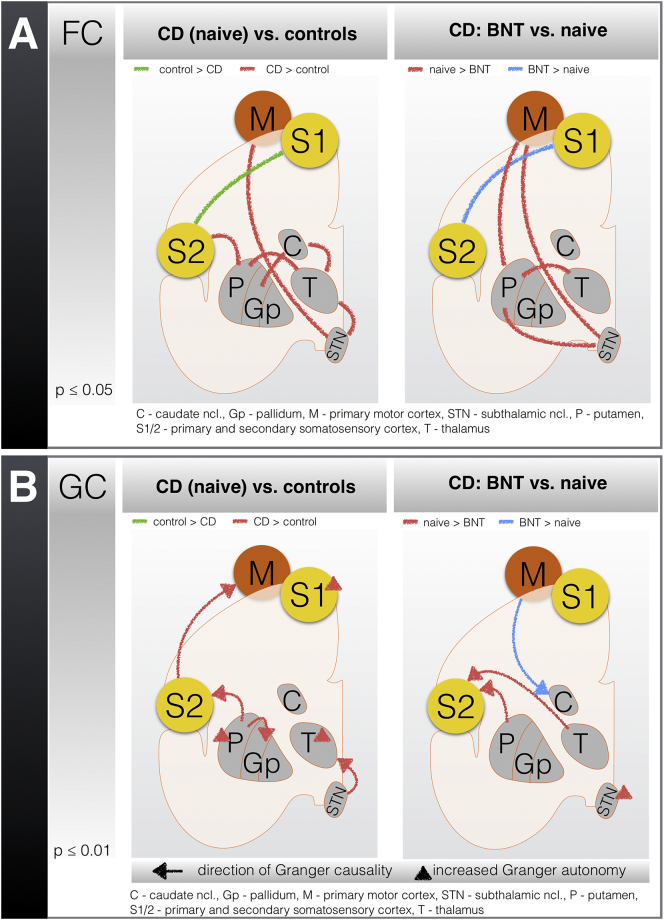
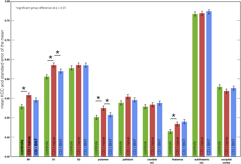
Methods: Using 3 T functional MR imaging along with advanced analysis techniques (functional connectivity, Granger causality, and regional homogeneity), we aimed to characterize brain activity in CD (17 CD patients vs. 17 controls) and to uncover the effects of BNT treatment (at 6 months).

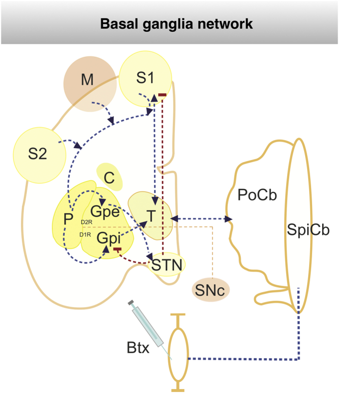
Results: In CD, we observed an increased information flow within the basal ganglia, the thalamus, and the sensorimotor cortex. In parallel, some of these structures became less responsive to regulating inputs. Furthermore, our results suggested an altered somatosensory integration. Following BNT administration, we noted a shift towards normal brain function in the CD patients, especially within the motor cortex, the somatosensory cortex, and the basal ganglia.

Conclusion: The changes in brain function and network activity in CD can be interpreted as related to the underlying cause, the effort to compensate or a mixture of both. Although BNT is applied in the last stage of the cortico-neuromuscular pathway, brain patterns are shifted towards those of healthy controls.

Keywords: Basal ganglia; Botulinum toxin (BNT); Cervical dystonia; Sensorimotor integration; Thalamus.


Figures

Fig. 1
Fig. 1
Schematic representation of botox treatment (BNT) in basalganglia network. The well characterized basal-ganglia loops, thalamo-cortical and cerebellar-thalamic loops are illustrated. In particular the main routes of the striato-fugal connections are displayed: the direct pathway with D2R-expressing striatal MSNs innervating the GPe and the indirect pathway with D1R-expressing striatal MSNs projecting to the GPi and STN. Abbreviations: Btx - Botoliumtoxin, C - caudate ncl, D1R – dopamin receptor 1, D2R – dopamine receptor 2, GPi - internal pallidum, Gpe - external pallidum, M - primary cortex, MSN - medium spiny neurons, STN - subthalamic ncl, PoCb - pontocerebellum, P - putamen, S1/2 - primary and secondary somatosensory cortex, SNC - substantia nigra pars compacta, SpCB - spinocerebellum, T - thalamus, dashed red lines: mainly inhibition connection, dashed blue lines: activating connection, dashed orange lines: dopaminergic innervation. (For interpretation of the references to color in this figure legend, the reader is referred to the web version of this article.)
Fig. 2
Fig. 2
Results from the functional (A) and effective (B) connectivity analyses of patients with cervical dystonia (CD) and the effects of treatment with botulinum toxin (BNT). Schematic representation of (A) functional connectivity (FC) and (B) Granger causality (GC) in resting-state fMRI from (left panels) the controls compared with the patients with CD (naive to treatment with BNT) and (right panels) the patients with CD who were treated without (naive) and with BNT (6 months). The results for the comparisons between patients with CD and controls (independent samples) and between patients with CD before and after BNT treatment (dependent samples) were corrected for multiple comparisons using a Bonferroni/FWE correction. In the figure, the lines indicate increased connectivity between specific brain regions; in GC, the direction of the arrow indicates the direction of the effective connectivity. A / left panel: in controls, FC between S1/S2 is increased; in patients with CD, increased connectivity within the BG, the thalamus, the motor cortex, and S2 is observed. A / right panel: the connectivity between S1/S2 is increased in patients with CD following BNT treatment; before the BNT application, the FC between the putamen and the thalamus, the STN, and the motor cortex is increased. B / left panel: in patients with CD, increased coupling of the putamen towards the pallidum and S2, S2 towards the motor cortex, and STN towards the thalamus is observed; increased autonomy in the pallidum and the thalamus is also detected. B / right panel: increased coupling of the motor cortex towards the caudate nucleus is observed in patients with CD following the BNT application; before the BNT application, increased GC exists between the putamen and the thalamus towards S2 and increased autonomy of the STN.
Fig. 3
Fig. 3
Regional homogeneity analysis in cervical dystonia (CD) and effects of treatment with botulinum toxin (BNT). Kendall's coefficient of concordance (KCC) and standard error of the mean of the defined ROIs in the primary motor cortex (M1), the primary and secondary somatosensory cortex (S1 / S2), the BG, and the thalamus in the controls and the CD patients without (naive) and with BNT (6 month) treatment. In CD (naive) KCC values were significantly increased (p ≤ 0.01, 2-samples t-test: red vs. green) in M1, S1, putamen, and thalamus (compared to healthy controls). With BNT treatment KCC values in CD were again significantly decreased (p ≤ 0.01, paired t-test: blue vs. red) in S1 and putamen. (For interpretation of the references to color in this figure legend, the reader is referred to the web version of this article.)

References

    1. Abbruzzese G., Berardelli A. Sensorimotor integration in movement disorders. Mov. Disord. 2003;18(3):231–240. - PubMed
    1. Albanese A., Bhatia K., Bressman S.B., Delong M.R., Fahn S., Fung V.S.C.…Teller J.K. Phenomenology and classification of dystonia: a consensus update. Mov. Disord. 2013;28(7):863–873. - PMC - PubMed
    1. Albin R.L., Young A.B., Penney J.B. The functional anatomy of basal ganglia disorders. Trends Neurosci. 1989;12(10):366–375. - PubMed
    1. Antelmi E., Erro R., Rocchi L., Liguori R., Tinazzi M., Di Stasio F.…Bhatia K.P. Neurophysiological correlates of abnormal somatosensory temporal discrimination in dystonia. Mov. Disord. 2017;32(1):141–148. - PubMed
    1. Bara-Jimenez W., Shelton P., Hallett M. Spatial discrimination is abnormal in focal hand dystonia. Neurology. 2000;55(12):1869–1873. - PubMed

Publication types

MeSH terms