. 2019 Apr 4;104(4):767-773.
doi: 10.1016/j.ajhg.2019.03.001. Epub 2019 Mar 28.

Truncating Mutations in UBAP1 Cause Hereditary Spastic Paraplegia

Affiliations

Truncating Mutations in UBAP1 Cause Hereditary Spastic Paraplegia

Mohammad Ali Farazi Fard et al. Am J Hum Genet. .

Erratum in

  • Truncating Mutations in UBAP1 Cause Hereditary Spastic Paraplegia.
    Farazi Fard MA, Rebelo AP, Buglo E, Nemati H, Dastsooz H, Gehweiler I, Reich S, Reichbauer J, Quintáns B, Ordóñez-Ugalde A, Cortese A, Courel S, Abreu L, Powell E, Danzi MC, Martuscelli NB, Bis-Brewer DM, Tao F, Zarei F, Habibzadeh P, Yavarian M, Modarresi F, Silawi M, Tabatabaei Z, Yousefi M, Farpour HR, Kessler C, Mangold E, Kobeleva X, Tournev I, Chamova T, Mueller AJ, Haack TB, Tarnopolsky M, Gan-Or Z, Rouleau GA, Synofzik M, Sobrido MJ, Jordanova A, Schüle R, Zuchner S, Faghihi MA. Farazi Fard MA, et al. Am J Hum Genet. 2019 Jun 6;104(6):1251. doi: 10.1016/j.ajhg.2019.05.009. Am J Hum Genet. 2019. PMID: 31173719 Free PMC article. No abstract available.

Abstract

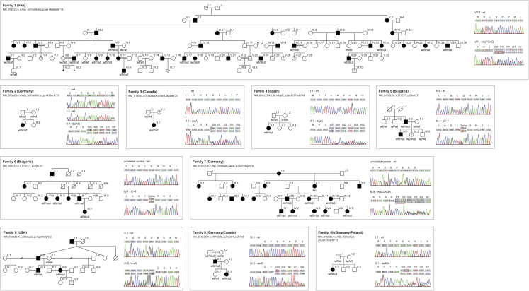
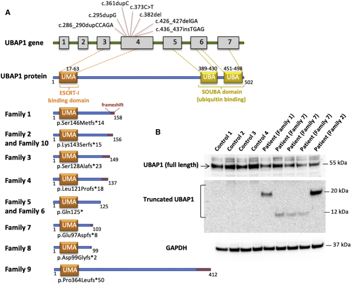
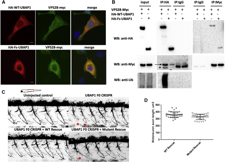
The diagnostic gap for rare neurodegenerative diseases is still considerable, despite continuous advances in gene identification. Many novel Mendelian genes have only been identified in a few families worldwide. Here we report the identification of an autosomal-dominant gene for hereditary spastic paraplegia (HSP) in 10 families that are of diverse geographic origin and whose affected members all carry unique truncating changes in a circumscript region of UBAP1 (ubiquitin-associated protein 1). HSP is a neurodegenerative disease characterized by progressive lower-limb spasticity and weakness, as well as frequent bladder dysfunction. At least 40% of affected persons are currently undiagnosed after exome sequencing. We identified pathological truncating variants in UBAP1 in affected persons from Iran, USA, Germany, Canada, Spain, and Bulgarian Roma. The genetic support ranges from linkage in the largest family (LOD = 8.3) to three confirmed de novo mutations. We show that mRNA in the fibroblasts of affected individuals escapes nonsense-mediated decay and thus leads to the expression of truncated proteins; in addition, concentrations of the full-length protein are reduced in comparison to those in controls. This suggests either a dominant-negative effect or haploinsufficiency. UBAP1 links endosomal trafficking to the ubiquitination machinery pathways that have been previously implicated in HSPs, and UBAP1 provides a bridge toward a more unified pathophysiology.

Keywords: animal model; endosomal trafficking; genetic diseases; hereditary spastic paraplegia; neurodegenerative diseases; spasticity; ubiquitination; zebrafish.


Figures

Figure 1
Figure 1
Pedigrees of HSP-Affected Families with UBAP1 Truncations All pedigrees suggest an autosomal-dominant or a de novo Mendelian trait. HSP-affected individuals are marked by filled symbols; individuals with unclear affection status are marked by a question mark. “mut” depicts the presence of a causative allele. Sanger traces exemplify the confirmation of variants detected via next-generation sequencing. The penetrance of truncating UBAP1 variants is reduced: individual F5-III.2 was subjectively unaffected at age 14 but showed brisk reflexes of lower limbs, indicating potential dysfunction of the corticospinal tract. The 80-year-old grandfather of the index case in family 9 (F9-III.1) was unfortunately not available for a neurological examination but was reported to be in good health and without any indication of a gait disturbance.
Figure 2
Figure 2
The Structure of UBAP1 and Mutations Carried by Affected Individuals (A) Schematic diagram showing all exons and UTRs of UBAP1 on the basis of gene model GenBank: NM_016525.4. The gray boxes represent the coding sequence of UBAP1. All variants occurred in exon 4 of UBAP1. The UMA protein domain includes amino acids 17–63, and two UBAs include amino acids 389–430 and 451–489. All truncations are listed below; the preserving of the UMA domain is clearly depicted, but there is loss of the two SOUBA domains. (B) Immunoblot analysis with an antibody recognizing amino acids 25–75 of UBAP1 shows a notable decrease in the amount of full-length UBAP1 in fibroblasts of the affected individuals from three different families when these cells are compared to control fibroblasts. Truncated UBAP1 of the predicted sizes was detected in fibroblasts from affected individuals but not in control fibroblasts.
Figure 3
Figure 3
Functional In Vitro and In Vivo Studies of Truncated UBAP1 (A) Immunostaining of U2OS cells transfected with HA-WT-UBAP1 HA-Fs-UBAP1 (p.Leu121Profs18) and VPS-28-Myc. Both wild-type and mutant UBAP1 co-localize with VPS28-Myc. (B) A co-immunoprecipitation assay shows protein-protein interaction between VPS28-Myc and both HA-WT-UBAP1 and HA-Fs-UBAP1. Ubiquitinated proteins co-immunoprecipitated with HA-WT-UBAP1 but not with HA-Fs-UBAP1. The arrow points to VPS28-Myc, and the asterisk bellow the arrow indicates the IgG band. (C) Motor-neuron axons in Tg(olig2DsRed) zebrafish embryos at 48 hpf. Embryos were injected with CRISPR Cas9 and sgRNAs against UBAP1; injection was supplemented with human RNA rescue of wild-type or truncated mutant UBAP1. Truncated and misshaped axons were more commonly observed with mutant hRNA rescue (indicated by asterisks). Scale bars represent 50 μm. The phenotypic difference between treated groups was evaluated by a Fisher exact test. Samples were assigned to either normal or affected categories on the basis of the presence of truncated and misshaped axons. The Fisher exact statistic value was determined to be 0.003; the result is significant at p < 0.005. Statistics describing normal versus affected phenotypes were calculated on the basis of the following sample sizes (number of embryos observed as having a phenotype). F0 CRISPR + wild-type hRNA rescue: normal = 11, affected = 1. F0 CRISPR + mutant hRNA rescue (family 4, p.Leu121Profs18), normal = 9, affected = 15. (D) Quantification of the individual motor-axon lengths. p values were calculated with a one-tailed Student’s t test.: p = 0.0008 and n = 9 (number of embryos in each experimental group; four axons were measured per embryo).

References

    1. Fink J.K. Hereditary spastic paraplegia: Clinico-pathologic features and emerging molecular mechanisms. Acta Neuropathol. 2013;126:307–328. - PMC - PubMed
    1. Parodi L., Fenu S., Stevanin G., Durr A. Hereditary spastic paraplegia: More than an upper motor neuron disease. Rev. Neurol. (Paris) 2017;173:352–360. - PubMed
    1. Schüle R., Wiethoff S., Martus P., Karle K.N., Otto S., Klebe S., Klimpe S., Gallenmüller C., Kurzwelly D., Henkel D. Hereditary spastic paraplegia: Clinicogenetic lessons from 608 patients. Ann. Neurol. 2016;79:646–658. - PubMed
    1. Bis-Brewer D.M., Züchner S. Perspectives on the genomics of HSP beyond Mendelian inheritance. Front. Neurol. 2018;9:958. - PMC - PubMed
    1. Li H., Durbin R. Fast and accurate short read alignment with Burrows-Wheeler transform. Bioinformatics. 2009;25:1754–1760. - PMC - PubMed

Publication types

MeSH terms