. 2019 Apr 4;104(4):721-730.
doi: 10.1016/j.ajhg.2019.02.016. Epub 2019 Mar 28.

Mutations in the Neuronal Vesicular SNARE VAMP2 Affect Synaptic Membrane Fusion and Impair Human Neurodevelopment

Collaborators, Affiliations

Mutations in the Neuronal Vesicular SNARE VAMP2 Affect Synaptic Membrane Fusion and Impair Human Neurodevelopment

Vincenzo Salpietro et al. Am J Hum Genet. .

Abstract

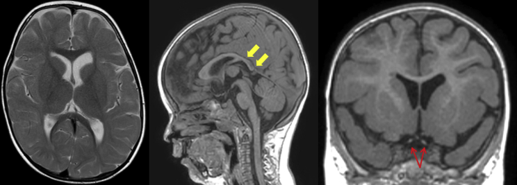
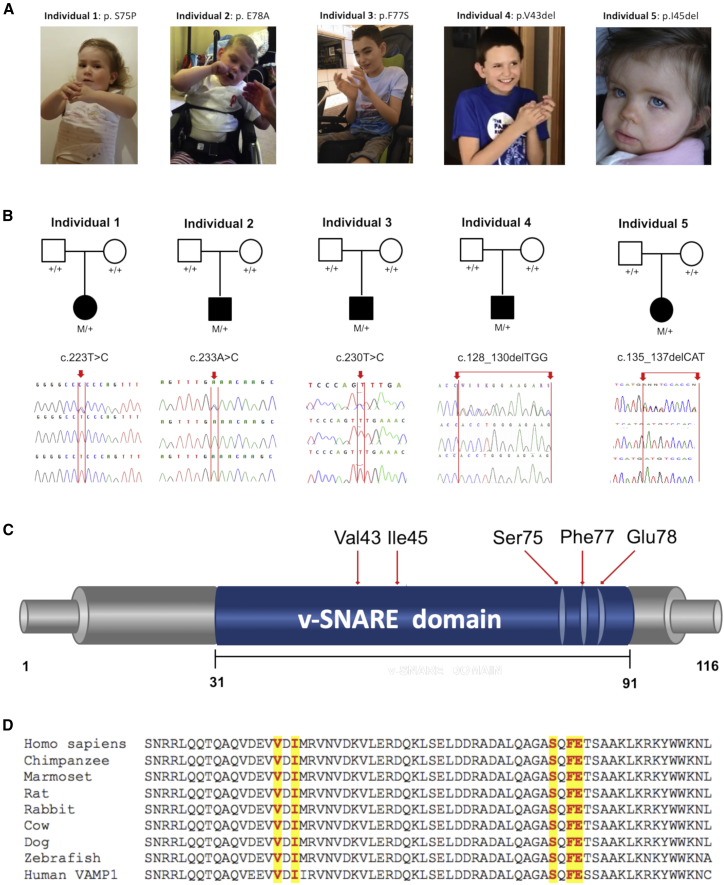
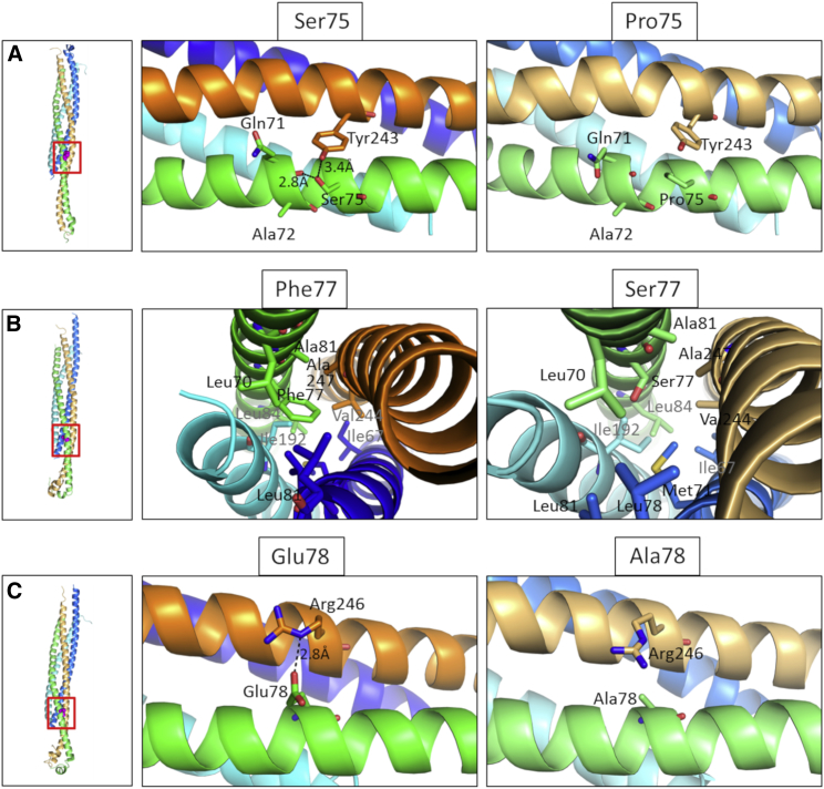
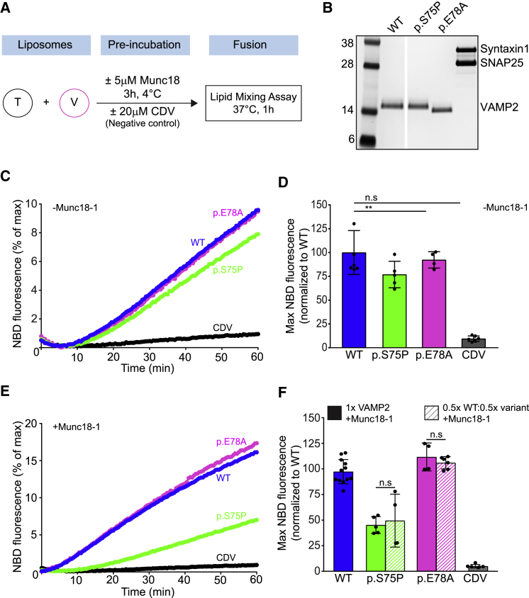
VAMP2 encodes the vesicular SNARE protein VAMP2 (also called synaptobrevin-2). Together with its partners syntaxin-1A and synaptosomal-associated protein 25 (SNAP25), VAMP2 mediates fusion of synaptic vesicles to release neurotransmitters. VAMP2 is essential for vesicular exocytosis and activity-dependent neurotransmitter release. Here, we report five heterozygous de novo mutations in VAMP2 in unrelated individuals presenting with a neurodevelopmental disorder characterized by axial hypotonia (which had been present since birth), intellectual disability, and autistic features. In total, we identified two single-amino-acid deletions and three non-synonymous variants affecting conserved residues within the C terminus of the VAMP2 SNARE motif. Affected individuals carrying de novo non-synonymous variants involving the C-terminal region presented a more severe phenotype with additional neurological features, including central visual impairment, hyperkinetic movement disorder, and epilepsy or electroencephalography abnormalities. Reconstituted fusion involving a lipid-mixing assay indicated impairment in vesicle fusion as one of the possible associated disease mechanisms. The genetic synaptopathy caused by VAMP2 de novo mutations highlights the key roles of this gene in human brain development and function.

Keywords: SNARE; VAMP2; autism; epilepsy; movement disorders; neurodevelopmental disorders; neuronal exocytosis; synaptobrevin; synaptopathy; vesicle fusion.


Figures

Figure 1
Figure 1
Brain MRI Scan of Individual 1, Who Harbors a De Novo VAMP2 p.Ser75Pro Variant, at the Age of 2 Years The panel shows axial T2-weighted, sagittal T1-weighted, and coronal T1-weighted MR images. There is some generalized delay in the maturation of myelin and a reduced volume of the cerebral white matter posteriorly. (Yellow arrows show a posteriorly slender corpus callosum.) The optic nerves and chiasm are hypoplastic (red arrows).
Figure 2
Figure 2
VAMP2 Intragenic De Novo Variants Identified in This Study (A) Individuals carrying de novo VAMP2 intragenic variants; note the hand stereotypies. (B) Sanger sequences of five kindreds with de novo VAMP2 intragenic variants. Chromatograms of individuals 1–5 and their parents confirm the de-novo occurrence of the VAMP2 variants in all cases. M/+ denotes the indicated VAMP2 variant in the heterozygous state, and +/+ denotes homozygous wild-type sequence. Mutant bases in the probands are indicated by a red arrow. (C) Schematic depiction of the human VAMP2 protein (GenBank: NP_055047.2) indicating the positions of the variants identified in this study. (D) Multiple alignment showing complete conservation across species and VAMP1 homolog (GenBank: NP_055046.1) of the residues affected by the variants identified in this study (these variants are highlighted in yellow). Human VAMP2 (GenBank: NP_055047.2), chimpanzee VAMP2 (UniProt: JAA33755.1), marmoset VAMP2 (UniProt: JAB33896.1), rat VAMP2 (NP_036795.1), rabbit VAMP2 (XP_008268978.1), cow VAMP2 (GenBank: NP_776908.1), dog VAMP2 (GenBank: XP_005620068.1), zebrafish VAMP2 (GenBank: NP_956299.1).
Figure 3
Figure 3
Molecular Modeling of the Identified De Novo VAMP2 Non-Synonymous Variants Comparison between the p.Ser75Pro (A), p.Phe77Ser (B), and p.Glu78Ala (C) mutant conformation within the SNARE complex (left panel, red square). The wild-type conformation is shown in the middle panel, and the mutated residues are shown in the right panel. Variant p.Ser75Pro causes the loss of two hydrogen bonds, one interchain between Ser75 of VAMP2 and Tyr243 of STX1A and one intrachain between Ser75 and Gln71; variant p.Phe77Ser introduces a hydrophilic residue in an otherwise hydrophobic region; and variant p.Glu78Ala causes the loss of a hydrogen bond between Glu78 of VAMP2 and Arg246 of STX1A. Modeling of the VAMP2 ectodomain (green for WT, light green for mutants) in complex with STX1A (orange for WT, light orange for mutants) and Snap25 (blue and cyan for WT, marine and aquamarine for mutants); configurations are as seen 100 ns into the molecular dynamic simulation. The complexes were modeled from the humanized 3HD7 complex. Water molecules and ions are not shown.
Figure 4
Figure 4
Disease-Associated VAMP2 Variants Result in Reduced Fusion Rates (A) Scheme showing the liposome fusion assay. (B) The SDS-PAGE and Coomassie-stained gel image of VAMP2 WT, VAMP2 disease-associated variants (p.Ser75Pro [p.Glu78Ala]), and t-SNARE (syntaxin 1 and SNAP25) reconstitution into donor v- and acceptor t-liposomes, respectively. (C) Line graphs showing the average basal (without Munc18-1) increase that occurs in NBD fluorescence as a result of fusion between the v-liposome and t-SNARE liposomes carrying WT or VAMP2 disease variants (p.Ser75Pro [p.Glu78Ala]). Liposome fusion reaction in the presence of CDV was used as negative control. (D) Basal fusion quantification, normalized to WT, at the endpoint (60 min) as described in (C). (E) Line graphs of liposome fusion reaction as in (C), in the presence of 5 μM Munc18-1. (F) Endpoint fusion quantification, normalized to WT, (60 min) of experiment as described in (E). Bar graphs also showed endpoint quantification of a similar experiment that used a v-liposome that contained a mixture of WT and mutant VAMP2 proteins. Data were from at least four independent replicates and presented as means plus SD. p < 0.05; ∗∗ p < 0.01; ∗∗∗ p < 0.001; n.s., not significant (p > 0.05).

References

    1. Jahn R., Fasshauer D. Molecular machines governing exocytosis of synaptic vesicles. Nature. 2012;490:201–207. - PMC - PubMed
    1. Hu C., Ahmed M., Melia T.J., Söllner T.H., Mayer T., Rothman J.E. Fusion of cells by flipped SNAREs. Science. 2003;300:1745–1749. - PubMed
    1. Chen Y.A., Scales S.J., Patel S.M., Doung Y.C., Scheller R.H. SNARE complex formation is triggered by Ca2+ and drives membrane fusion. Cell. 1999;97:165–174. - PubMed
    1. Li F., Kümmel D., Coleman J., Reinisch K.M., Rothman J.E., Pincet F. A half-zippered SNARE complex represents a functional intermediate in membrane fusion. J. Am. Chem. Soc. 2014;136:3456–3464. - PMC - PubMed
    1. Gao Y., Zorman S., Gundersen G., Xi Z., Ma L., Sirinakis G., Rothman J.E., Zhang Y. Single reconstituted neuronal SNARE complexes zipper in three distinct stages. Science. 2012;337:1340–1343. - PMC - PubMed

Publication types

MeSH terms