. 2019 Dec;70(6):2021-2031.e1.
doi: 10.1016/j.jvs.2018.12.022. Epub 2019 Mar 28.

The myristoylated alanine-rich C kinase substrate differentially regulates kinase interacting with stathmin in vascular smooth muscle and endothelial cells and potentiates intimal hyperplasia formation

Affiliations

The myristoylated alanine-rich C kinase substrate differentially regulates kinase interacting with stathmin in vascular smooth muscle and endothelial cells and potentiates intimal hyperplasia formation

Dan Yu et al. J Vasc Surg. 2019 Dec.

Abstract

Objective: Restenosis limits the durability of all cardiovascular reconstructions. Vascular smooth muscle cell (VSMC) proliferation drives this process, but an intact, functional endothelium is necessary for vessel patency. Current strategies to prevent restenosis employ antiproliferative agents that affect both VSMCs and endothelial cells (ECs). Knockdown of the myristoylated alanine-rich C kinase substrate (MARCKS) arrests VSMC proliferation and paradoxically potentiates EC proliferation. MARCKS knockdown decreases expression of the kinase interacting with stathmin (KIS), increasing p27kip1 expression, arresting VSMC proliferation. Here, we seek to determine how MARCKS influences KIS protein expression in these two cell types.

Methods: Primary human coronary artery VSMCs and ECs were used for in vitro experiments. MARCKS was depleted by transfection with small interfering RNA. Messenger RNA was quantitated with the real-time reverse transcription polymerase chain reaction. Protein expression was determined by Western blot analysis. Ubiquitination was determined with immunoprecipitation. MARCKS and KIS binding was assessed with co-immunoprecipitation. Intimal hyperplasia was induced in CL57/B6 mice with a femoral artery wire injury. MARCKS was knocked down in vivo by application of 10 μM of small interfering RNA targeting MARCKS suspended in 30% Pluronic F-127 gel. Intimal hyperplasia formation was assessed by measurement of the intimal thickness on cross sections of the injured artery. Re-endothelialization was determined by quantitating the binding of Evans blue dye to the injured artery.

Results: MARCKS knockdown did not affect KIS messenger RNA expression in either cell type. In the presence of cycloheximide, MARCKS knockdown in VSMCs decreased KIS protein stability but had no effect in ECs. The effect of MARCKS knockdown on KIS stability was abrogated by the 26s proteasome inhibitor MG-132. MARCKS binds to KIS in VSMCs but not in ECs. MARCKS knockdown significantly increased the level of ubiquitinated KIS in VSMCs but not in ECs. MARCKS knockdown in vivo resulted in decreased KIS expression. Furthermore, MARCKS knockdown in vivo resulted in decreased 5-ethynyl-2'-deoxyuridine integration and significantly reduced intimal thickening. MARCKS knockdown enhanced endothelial barrier function recovery 4 days after injury.

Conclusions: MARCKS differentially regulates the KIS protein stability in VSMCs and ECs. The difference in stability is due to differential ubiquitination of KIS in these two cell types. The differential interaction of MARCKS and KIS provides a possible explanation for the observed difference in ubiquitination. The effect of MARCKS knockdown on KIS expression persists in vivo, potentiates recovery of the endothelium, and abrogates intimal hyperplasia formation.

Keywords: Cell migration; Cell proliferation; KIS; MARCKS.


Figures

Figure 1.
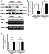
Figure 1.. MARCKS knockdown does not affect KIS mRNA expression in either VSMCs or ECs.
A. Representative Western blots showing successful inhibition of MARCKS protein expression at 72 hours after treatment with siMARCKS. Total cell lysates from human coronary artery VSMCs and human coronary artery ECs were prepared and analyzed with Western blot. GAPDH was used as loading control. B. Data from three independent experiments demonstrate that MARCKS knockdown decreases KIS expression in VSMCs but increases KIS expression in ECs. * denotes p<0.05. C. RT-PCR analysis of KIS mRNA expression in siRNA-treated cells as described in Figure 1A. MARCKS knockdown does not affect KIS mRNA expression. D. Quantitative real-time RT-PCR (qPCR) was used to quantify mRNA expression in VSMCs and ECs after treatment with siMARCKS or siControl. Values are presented as means ± standard deviation from three separate experiments. NS indicates no significant difference (p>0.05) between cells treated with siMARCKS and siControl.
Figure 2.
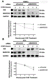
Figure 2.. MARCKS knockdown decreases KIS protein stability in VSMCs but increases KIS stability in ECs.
Human coronary artery VSMCs and ECs were grown in culture and transfected with either siMARCKS or siControl. The cells were then treated with 10 μg/ml cyclohexamide (CHX) to inhibit protein synthesis. Total cell lysates were prepared at time points indicated. KIS expression was determined with Western blot and GAPDH was used as loading control. A. In VSMCs, MARCKS KD decreased KIS expression at 4- and 8-hours after CHX treatment compared to time 0 (two-tailed Student’s t-test, p<0.05). B. In contrast, MARCKS knockdown did not affect KIS protein expression in ECs. Data are expressed as relative KIS/GAPDH ratios as compared to time 0. Values are presented as means ± standard deviation from three separate experiments.
Figure 3.
Figure 3.. MARCKS KD affects KIS ubiquitination.
A. Human coronary artery VSMCs were grown in culture and transfected with either siMARCKS or siControl. The cells were pretreated with MG-132 (10 nM) for 16 hours before incubated with cyclohexamide (CHX) (10 μg/ml) for protein degradation assay. Pre-treating cells with MG-132 (10 nM) abrogated the decreased KIS protein stability previously observed with MARCKS knockdown. B. Data are expressed as relative KIS/GAPDH level as compared with time 0. Data are presented as means ± standard deviations of three separate experiments. Statistical significance was determined by the two-tail Student’s t-test. No difference in KIS expression was detected in VSMCs with MARCKS knockdown in the presence of MG-132. C. KIS ubiquitination was analyzed in human coronary artery VSMCs and ECs. Cells were grown in culture and transfected with either siMARCKS or non-targeting, siControl. The cells were then treated with MG-132 to block the 26s proteasome function and prevent the degradation of ubiquitinated protein. Total cell lysates were prepared immunoprecipitated with an Ubiquitin Enrichment Kit (Pierce). The precipitate was blotted with anti-KIS (IP). To confirm equal target loading, the total cell lysate (TL) was blotted for GAPDH. D. MARCKS knockdown resulted in increased KIS ubiquitination in VSMCs, but paradoxically decreased KIS ubiquitination in ECs. Data are presented as means ± standard deviations of three separate experiments. Statistical significance was determined by the two-tail Student’s t-test. * denotes p<0.05).
Figure 4.
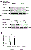
Figure 4.. MARCKS binds KIS in VSMCs but not in ECs.
A. Human coronary artery VSMCs and ECs were cultured sub-confluently and starved for 48 hours. Cells were then stimulated with 20% fetal bovine serum (FBS) for 10 min. Total cell lysates were analyzed with Western blot to determine the expression of MARKCS and KIS. GAPDH was used as loading control. B. MARCKS protein was immunoprecipitated (IP) with anti-MARCKS antibody from the total cell lysate then immunoblotted (IB) with anti-KIS antibody. The precipitate was also blotted with anti-MARCKS antibody to demonstrate equal input loading. C. MARCKS coprecipitates with KIS in VSMCs, but not ECs. This relationship was potentiated with mitogen (serum) exposure in VSMCs.
Figure 5.
Figure 5.. MARCKS knockdown prevents intima hyperplasia formation in vivo.
The mouse femoral artery wire injury model was used to generate intimal hyperplasia formation. The animals were subjected to bilateral femoral artery wire injury. At the time of surgery, the external surface of the artery was coated with either siControl or siMARCKS (10 μM) in a 30% pluronic gel solution. Animals were euthanized, and the femoral arteries were processed for histology at 6- and 12-weeks after injury. The sectioned femoral arteries were stained with hematoxylin and eosin and counter stained with Verhoeff-Van Gieson stain (VVG) to demonstrate the border between the intima and the media (arrows). A and B. At 6 weeks after injury, vessels treated with siControl had formed more intimal hyperplasia (Fig 5A) than those treated with siMARCKS (Fig 5B). C. The intima-to-media ratio (I/M ratio) was 1.16 ± 0.57 for vessels treated with siControl and 0.09 ± 0.02 for vessels treated with siMARCKS. Data are presented as means ± standard deviations for three independent experiments. * denotes p<0.05 as determined by the two-tailed Student-t test. Scale bars = 100 μm.
Figure 6.
Figure 6.. MARCKS knockdown decreases time to return of endothelial integrity after wire injury.
The mouse femoral artery wire injury model was used to generate an endothelial injury. The animals were subjected to bilateral femoral artery wire injury. At the time of surgery, the external surface of the artery was coated with either siControl or siMARCKS (10 μM in a 30% pluronic gel solution. At the time of euthanasia, the animals were perfused with 0.3% Evans Blue dye. A. Evans Blue dye is excluded from vessels with an intact endothelium and thus there is the minimal staining in the uninjured femoral artery. Wire injury disrupted the barrier function of the endothelium allowing the dye to penetrate the arterial wall and stain it blue. B and C. Four days after injury, vessels treated with siControl exhibited greater Evans Blue staining than vessels treated with siMARCKS. D. Evans Blue staining was further quantitated by measuring the amount of Evans Blue (ng) bound to the vessel and normalized to total vessel weight. Vessels treated with siControl bound significantly more Evans Blue (297 ±15.0 ng/mg) than vessels treated with siMARCKS (171 ± 37.5 ng/mg, p<0.05). Data are presented as means ± standard deviations for three independent experiments. * denotes p<0.05 as determined by the two-tailed Student-t test. Scale bars = 500 μm.
Figure 7.
Figure 7.. KIS protein expression and VSMC proliferation are attenuated by MARCKS knockdown.
The mouse femoral artery wire injury model of intimal hyperplasia was used to evaluate in vivo effects of MARCKS knockdown on VSMC proliferation and KIS expression. The animals were subjected to bilateral femoral artery wire injury. At the time of surgery, the external surface of the artery was coated with either siControl or siMARCKS (10 μM) in a 30% pluronic gel solution. Animals were euthanized 7 days after injury. A and B. Treatment with siMARCKS greatly reduced the amount of MARCKS protein expression throughout the vessel wall. C and D. MARCKS knockdown resulted in decreased KIS expression throughout the arterial wall. E and F. MARCKS knockdown reduced the vascular proliferative response to injury as measured by EdU incorporation. G-J. The vessels were counterstained for α-smc actin, and DAPI. K and L. Images for KIS, EdU, α-smc actin, and DAPI demonstrate colocalization of KIS and DAPI in vessels treated with siControl, but not siMARCKS. M. Treatment with siMARCKS resulted in significantly less KIS expressed in the vessel wall compared to treatment with siControl (1.47 ± 0.15 /1,000 μm2 compared to 1.47 ± 0.15 /1,000 μm2, p<0.05). N. Treatment with siMARCKS significantly reduced the vascular proliferative response to injury as determined by EdU incorporation (0.33 ± 0.11 /1,000μm2 compared to 2.90 ± 0.92 /1,000 μm2, p<0.05). Data are presented as means ± standard deviations for three independent experiments. * denotes p<0.05 as determined by the two-tailed Student’s-t test. Scale bars = 10 μm.

Comment in

  • Invited commentary.
    Ruddy JM. Ruddy JM. J Vasc Surg. 2019 Dec;70(6):2031-2032. doi: 10.1016/j.jvs.2018.12.021. J Vasc Surg. 2019. PMID: 31761108 No abstract available.

References

    1. Duda SH, Bosiers M, Lammer J, Scheinert D, Zeller T, Tielbeek A, et al. Sirolimus-eluting versus bare nitinol stent for obstructive superficial femoral artery disease: the SIROCCO II trial. J Vasc Interv Radiol. 2005;16(3):331–8. - PubMed
    1. Tepe G, Zeller T, Albrecht T, Heller S, Schwarzwalder U, Beregi JP, et al. Local delivery of paclitaxel to inhibit restenosis during angioplasty of the leg. N Engl J Med. 2008;358(7):689–99. - PubMed
    1. Holmes DR Jr., Leon MB, Moses JW, Popma JJ, Cutlip D, Fitzgerald PJ, et al. Analysis of 1-year clinical outcomes in the SIRIUS trial: a randomized trial of a sirolimus-eluting stent versus a standard stent in patients at high risk for coronary restenosis. Circulation. 2004;109(5):634–40. - PubMed
    1. Finn AV, Joner M, Nakazawa G, Kolodgie F, Newell J, John MC, et al. Pathological correlates of late drug-eluting stent thrombosis: strut coverage as a marker of endothelialization. Circulation. 2007; 115(18):2435–41. - PubMed
    1. Hayashi S, Yamamoto A, You F, Yamashita K, Ikegame Y, Tawada M, et al. The stenteluting drugs sirolimus and paclitaxel suppress healing of the endothelium by induction of autophagy. Am J Pathol. 2009;175(5):2226–34. - PMC - PubMed

Publication types

MeSH terms