. 2019 Apr 9;27(2):334-342.e10.
doi: 10.1016/j.celrep.2019.02.055. Epub 2019 Mar 28.

A Ubiquitous Platform for Bacterial Nanotube Biogenesis

Affiliations

A Ubiquitous Platform for Bacterial Nanotube Biogenesis

Saurabh Bhattacharya et al. Cell Rep. .

Abstract

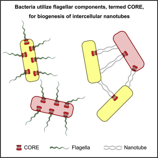
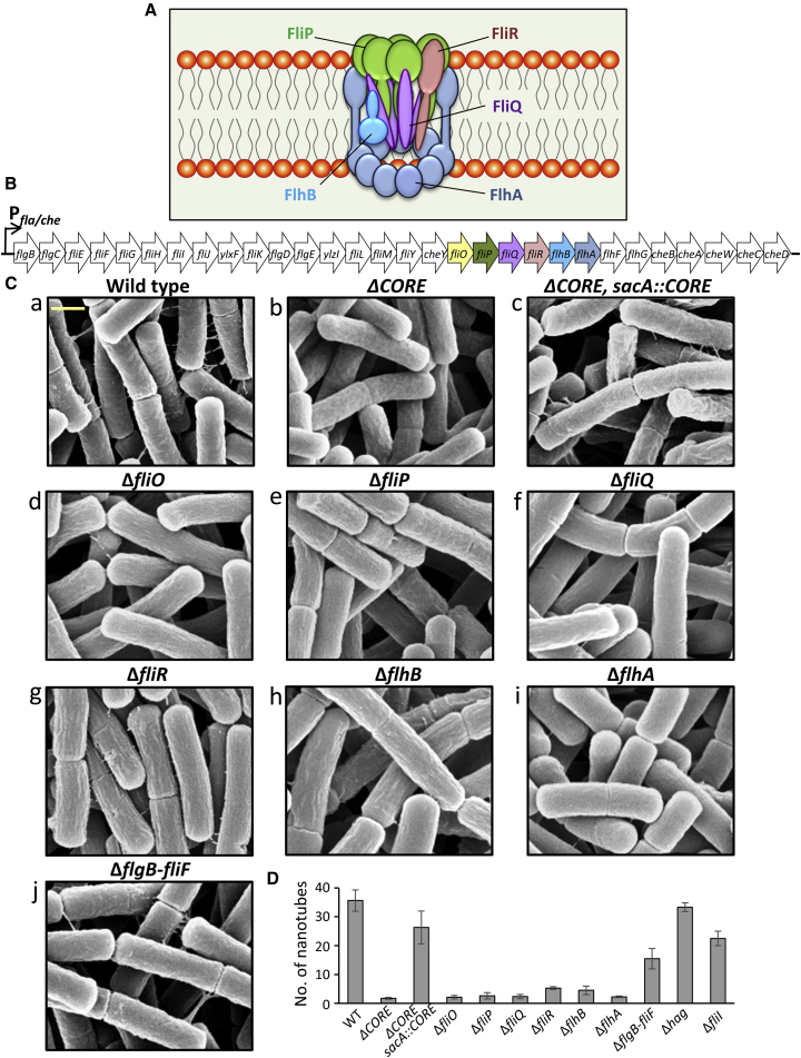
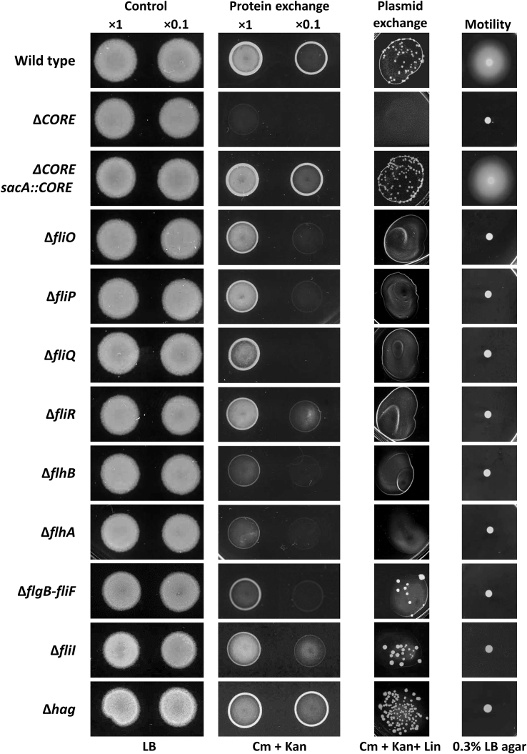
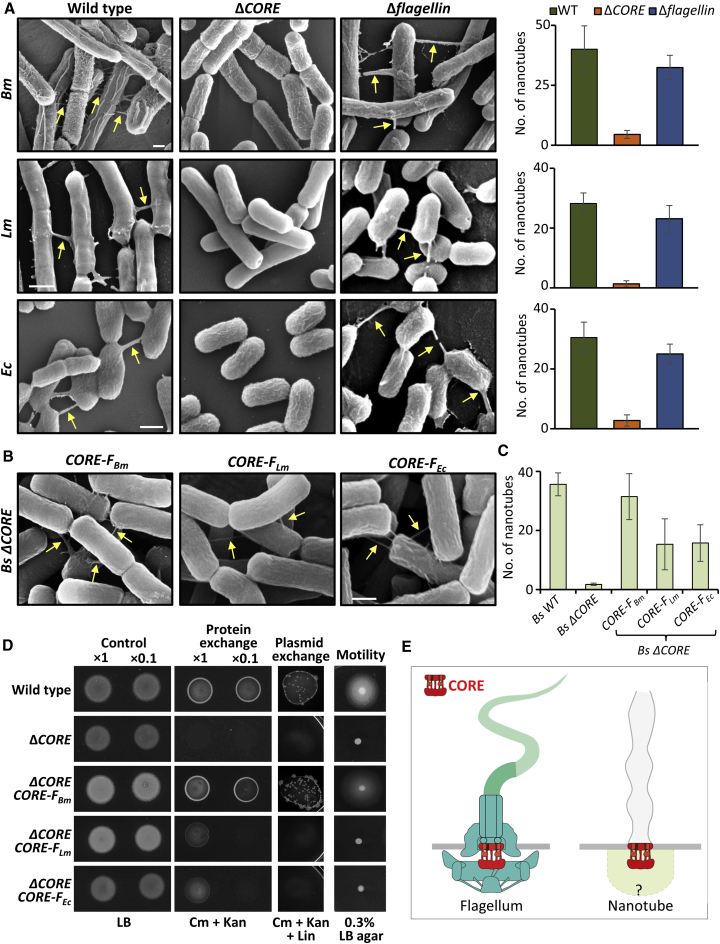
We have previously described the existence of membranous nanotubes, bridging adjacent bacteria, facilitating intercellular trafficking of nutrients, cytoplasmic proteins, and even plasmids, yet components enabling their biogenesis remain elusive. Here we reveal the identity of a molecular apparatus providing a platform for nanotube biogenesis. Using Bacillus subtilis (Bs), we demonstrate that conserved components of the flagellar export apparatus (FliO, FliP, FliQ, FliR, FlhB, and FlhA), designated CORE, dually serve for flagellum and nanotube assembly. Mutants lacking CORE genes, but not other flagellar components, are deficient in both nanotube production and the associated intercellular molecular trafficking. In accord, CORE components are located at sites of nanotube emergence. Deleting COREs of distinct species established that CORE-mediated nanotube formation is widespread. Furthermore, exogenous COREs from diverse species could restore nanotube generation and functionality in Bs lacking endogenous CORE. Our results demonstrate that the CORE-derived nanotube is a ubiquitous organelle that facilitates intercellular molecular trade across the bacterial kingdom.

Keywords: Bacillus subtilis; bacterial communication; bacterial community; contact-dependent molecular exchange; flagella type III secretion system; flagellar export apparatus; nanotubes.


Figures

None
Graphical abstract
Figure 1
Figure 1
Bs CORE Mutants Are Impaired in Nanotube Formation (A) Schematic illustration of the flagellar CORE apparatus on the basis of Fukumura et al. (2017) and Kuhlen et al. (2018). The CORE consists of FliP, FliQ, FliR, FlhB, and FlhA (5:4:1:1:9) transmembrane proteins and the chaperone FliO (not shown), which only transiently associates with the CORE complex. (B) A map depicting the fla/che operon of Bs, encoding the components required for flagellar basal body formation. Genes encoding the CORE proteins are highlighted with the same color code as in (A). (C) The indicated Bs CORE mutant strains were visualized using XHR-SEM to monitor the formation of intercellular nanotubes. Strains were grown to the mid-logarithmic phase, spotted onto EM grids, incubated on LB agar plates for 4 h at 37°C, and visualized using XHR-SEM. Scale bar represents 500 nm. (D) Quantification of the average number of nanotubes displayed per 50 cells by the indicated Bs mutant strains following XHR-SEM analysis described in (C). Shown are average values and SD of at least three independent experiments (n ≥ 200 for each strain). See also Figure S1.
Figure 2
Figure 2
Bs CORE Mutants Are Deficient in Molecular Exchange Assessing molecular exchange in CORE mutants. For protein exchange assay, pairs of a donor (SB463: amyE::Phyper-spank-cat-spec) (CmR, SpecR) and a recipient (SB513: amyE::Phyper-spank-gfp-kan) (KanR) parental strains (wild-type) were used. The investigated mutants harbor the corresponding genotypes and carry the indicated null mutation in both donor and recipient strains. Donor and recipient strains were mixed in 1:1 ratio (at two concentrations, 1× and 0.1×) and incubated in LB supplemented with 1 mM IPTG for 4 h at 37°C with gentle shaking. Equal numbers of cells were then spotted onto LB agar (control) and LB agar containing chloramphenicol (Cm) and kanamycin (Kan) (protein exchange) and photographed after 18 h. For plasmid exchange assay, pairs of a donor (GD110: amyE::Phyper-spank-cat-spec, pHB201/cat, erm) (CmR, SpecR, MlsR) and a recipient (SB513: amyE::Phyper-spank-gfp-kan) (KanR) parental strains (wild-type) were used, with the investigated mutants harbor in addition the indicated null mutation in both donor and recipient strains. Cells were mixed in 1:1 ratio (concentration 1×), processed as described for protein exchange, and spotted onto LB agar containing Cm, Kan, and lincomycin (Lin) (plasmid exchange). Cells were incubated at 37°C, and colonies were photographed after 36 h of incubation. For motility assay, wild-type (PY79) and the indicated mutant strains were grown to the mid-logarithmic phase and spotted onto LB plates containing 0.3% agar and photographed after 7 h of incubation at 37°C (motility). See also Figures S1 and S2.
Figure 3
Figure 3
FliP Localizes to the Base of Nanotubes Cells expressing HA-tagged FliP (SH93: amyE::Phyper-spank-fliP2xHA, sacA::Phyper-spank-ymdB, A and B, or SH110: amyE::Phyper-spank-fliP2xHA, sacA::Phyper-spank-ymdB, Δmbl, C and D) were spotted onto EM grids and subjected to immuno-gold XHR-SEM, using primary antibodies against HA and secondary gold-conjugated antibodies. Samples were not coated before observation. Examples of FliP2xHA localization (white dots) at the base of nanotubes are presented (indicated by arrows). Shown are XHR-SEM images that were acquired using TLD-SE (through-lens detector-secondary electron) for nanotubes visualization and vCD (low-kV high-contrast backscattered detector) for gold particle detection, as well as overlay of both images. Schematic below depicts the interpretive cell layout and highlights the nanotube region with gold signal (dashed box) captured by XHR-SEM. Scale bars represent 200 nm. See also Figures S3 and S4.
Figure 4
Figure 4
CORE Is a Widespread Complex with Conserved Functions across Species (A) Wild-type Bm (OS2), Lm (10403S), and Ec (MG1655) and their corresponding mutant strains lacking CORE (ΔCORE) or gene encoding flagellin (Δflagellin) were grown to the mid-logarithmic phase, spotted onto EM grids followed by incubation on LB agar plates for 4 h at 37°C, and visualized using XHR-SEM. Arrows indicate nanotubes. Scale bar represents 500 nm. Right: quantification of the average number of nanotubes displayed per 50 cells by the indicated strains following XHR-SEM analysis. Shown are average values and SD of at least three independent experiments (n ≥ 200 for each strain). (B) Cells of BsCORE) complemented with Bm flagellar CORE (CORE-FBm), Lm flagellar CORE (CORE-FLm), and Ec flagellar CORE (CORE-FEc) were processed as in (A) and visualized using XHR-SEM. Arrows indicate nanotubes. Scale bar represents 500 nm. (C) Quantification of the average number of nanotubes displayed per 50 cells by the indicated strains following XHR-SEM analysis described in (B). Shown are average values and SD of at least three independent experiments (n ≥ 200 for each strain). (D) Assessing molecular exchange in Bs ΔCORE strain complemented with exogenous CORE genes. For protein exchange assay, pairs of a donor (SB463: amyE::Phyper-spank-cat-spec) (CmR, SpecR) and a recipient (SB513: amyE::Phyper-spank-gfp-kan) (KanR) parental strains (wild-type) were used. The investigated strains ΔCORE and ΔCORE complemented with CORE-FBm, CORE-FLm, and CORE-FEc (controlled by Bs Pfla/che promoter) harbor the corresponding genotypes of donor and recipient. Donor and recipient strains were mixed in 1:1 ratio (at two concentrations, 1× and 0.1×) and incubated in LB supplemented with 1 mM IPTG for 4 h at 37°C with gentle shaking. Equal numbers of cells were then spotted onto LB agar (control) and LB agar containing chloramphenicol (Cm) and kanamycin (Kan) (protein exchange) and photographed after 18 h. For plasmid exchange assay, pairs of a donor (GD110: amyE::Phyper-spank-cat-spec, pHB201/cat, erm) (CmR, SpecR, MlsR) and a recipient (SB513: amyE::Phyper-spank-gfp-kan) (KanR) parental strains (wild-type) were used. The investigated strains complemented with exogenous COREs harbor the corresponding genotypes of donor and recipient strains. Cells were mixed in 1:1 ratio (concentration 1×), processed as described for protein exchange, and spotted onto LB agar containing Cm, Kan, and lincomycin (Lin) (plasmid exchange). Cells were incubated at 37°C, and colonies were photographed after 36 h of incubation. For motility assay, wild-type (PY79) and the indicated strains were grown to mid-logarithmic phase, spotted onto LB plates containing 0.3% agar, and photographed after 7 h of incubation at 37°C (motility). (E) A schematic model depicting the modularity of CORE complexes in flagellum and nanotube. CORE-associated components of the nanotube basal body are missing. Flagellum structure was adapted from (Dietsche et al., 2016). See also Figure S5 and Tables S1 and S2.

References

    1. Abby S.S., Rocha E.P. The non-flagellar type III secretion system evolved from the bacterial flagellum and diversified into host-cell adapted systems. PLoS Genet. 2012;8:e1002983. - PMC - PubMed
    1. Allen R.D., Baumann P. Structure and arrangement of flagella in species of the genus Beneckea and Photobacterium fischeri. J. Bacteriol. 1971;107:295–302. - PMC - PubMed
    1. Argov T., Rabinovich L., Sigal N., Herskovits A.A. An effective counterselection system for Listeria monocytogenes and its use to characterize the monocin genomic region of strain 10403S. Appl. Environ. Microbiol. 2017;83:e02927-16. - PMC - PubMed
    1. Baidya A.K., Bhattacharya S., Dubey G.P., Mamou G., Ben-Yehuda S. Bacterial nanotubes: a conduit for intercellular molecular trade. Curr. Opin. Microbiol. 2018;42:1–6. - PubMed
    1. Bejerano-Sagie M., Oppenheimer-Shaanan Y., Berlatzky I., Rouvinski A., Meyerovich M., Ben-Yehuda S. A checkpoint protein that scans the chromosome for damage at the start of sporulation in Bacillus subtilis. Cell. 2006;125:679–690. - PubMed

Publication types

MeSH terms

Substances

LinkOut - more resources