. 2019 May 8;27(5):986-998.
doi: 10.1016/j.ymthe.2019.03.007. Epub 2019 Mar 15.

Clinically Relevant Correction of Recessive Dystrophic Epidermolysis Bullosa by Dual sgRNA CRISPR/Cas9-Mediated Gene Editing

Affiliations

Clinically Relevant Correction of Recessive Dystrophic Epidermolysis Bullosa by Dual sgRNA CRISPR/Cas9-Mediated Gene Editing

Jose Bonafont et al. Mol Ther. .

Abstract

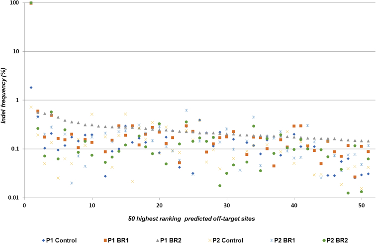
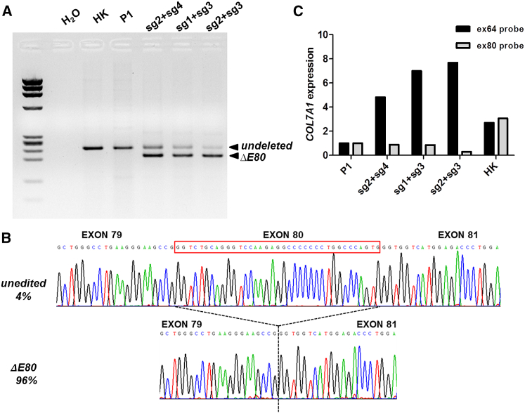
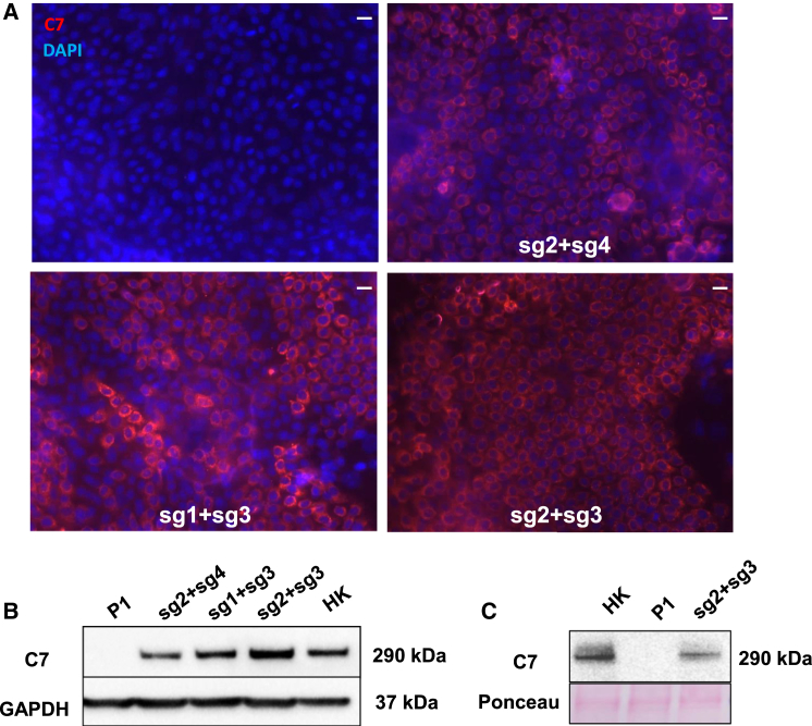
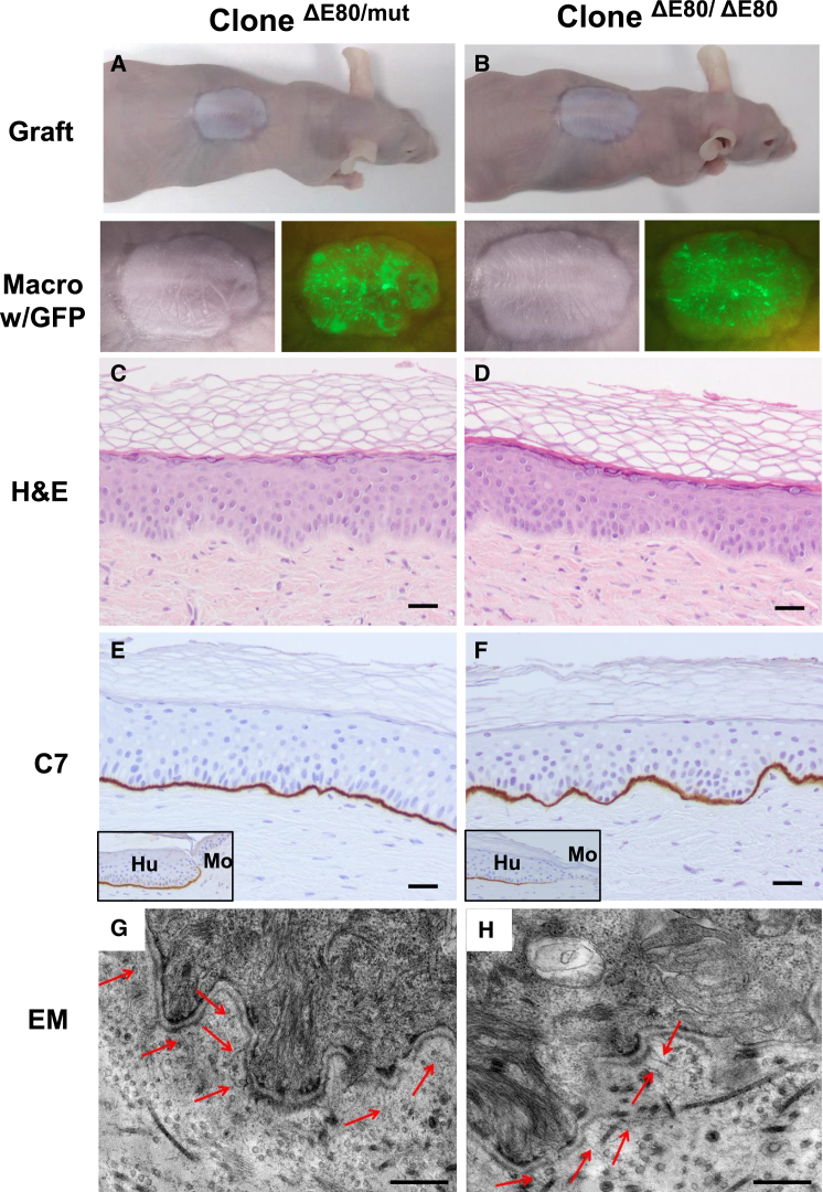
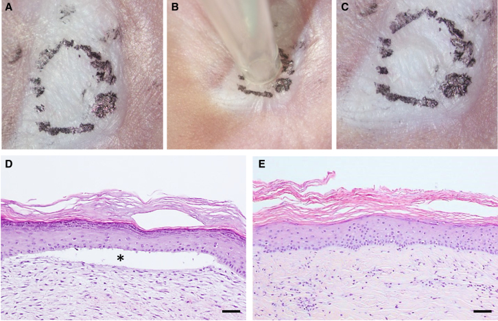
Gene editing constitutes a novel approach for precisely correcting disease-causing gene mutations. Frameshift mutations in COL7A1 causing recessive dystrophic epidermolysis bullosa are amenable to open reading frame restoration by non-homologous end joining repair-based approaches. Efficient targeted deletion of faulty COL7A1 exons in polyclonal patient keratinocytes would enable the translation of this therapeutic strategy to the clinic. In this study, using a dual single-guide RNA (sgRNA)-guided Cas9 nuclease delivered as a ribonucleoprotein complex through electroporation, we have achieved very efficient targeted deletion of COL7A1 exon 80 in recessive dystrophic epidermolysis bullosa (RDEB) patient keratinocytes carrying a highly prevalent frameshift mutation. This ex vivo non-viral approach rendered a large proportion of corrected cells producing a functional collagen VII variant. The effective targeting of the epidermal stem cell population enabled long-term regeneration of a properly adhesive skin upon grafting onto immunodeficient mice. A safety assessment by next-generation sequencing (NGS) analysis of potential off-target sites did not reveal any unintended nuclease activity. Our strategy could potentially be extended to a large number of COL7A1 mutation-bearing exons within the long collagenous domain of this gene, opening the way to precision medicine for RDEB.

Keywords: CRISPR/Cas9; epidermal stem cells; epidermolysis bullosa; gene therapy.


Figures

Figure 1
Figure 1
COL7A1 E80 Dual RNA Guide CRISPR/Cas9 Deletion Strategy (A) Scheme of the strategy. Single CRISPR RNA guides were designed to induce Cas9-mediated DNA double-strand breaks within selected COL7A1 intron sequences flanking exon 80 (U-Guides and D-Guides). NHEJ repair leads to intron-intron rejoining with concomitant E80 deletion and restoration of COL7A1 reading frame and truncated C7 expression. (B) CRISPR guide design. sgRNA (sgRNAs 1, 2, 3, and 4) sequences and alignment to E80-flanking sequences E80 are shown. The protospacer adjacent motif (PAM) is indicated in a darker color. (C) Predicted amplicon sizes for E80 deletions corresponding to each sgRNA pair combination. (D) PCR analysis of genomic DNA from RDEB keratinocytes from patient P1 treated with the RNP complexes containing the different sgRNA pair combinations. The sg2 + sg3 pair yielded the highest proportion of excised E80 according to the intensity of the lower 440-bp band. Deletion ratios assessed by densitometry are shown in each lane. “A” and “B” refer to the two different electroporation conditions tested.
Figure 2
Figure 2
Indel Spectrum in COL7A1 E80 Region Sequences (Sanger) listed from higher to lower frequencies. 50% of rejoining events corresponded to the fusion between E80-flanking cutting sites plus the addition of a T (Δ55 + insT). 95.14% of all alleles had been edited and 84.16% presented E80 deletion.
Figure 3
Figure 3
NGS Assessment for the Presence of Indels in Potential Off-Target Sites PCR amplicons for 244 predicted off-target sites sequenced with at least 1,000× depth, as well as the on-target region, were analyzed (x axis, putative off-target sites arranged according to the frequency of sequence variation). The 50 highest ranking sites are represented. The first point corresponds to the on-target site (y axis, percentage of reads containing sequence variations within a 6-bp window centered on the target site for each sgRNA).
Figure 4
Figure 4
Collagen VII mRNA Expression of ΔE80 Gene-Edited RDEB Keratinocytes (A) RT-PCR analysis of COL7A1 transcripts amplified with primers in exons 78–84. Wild-type/c.6527insC unedited transcripts produced a 240/241-bp band (non-modified, n-m) found in all RNA samples. A smaller 205-bp band corresponding to transcripts lacking exon 80 was detected in samples from edited cells. M, DNA Molecular Weight Marker IX (Sigma-Aldrich) molecular weight marker; HK, healthy human keratinocytes; P1, patient keratinocytes; H2O, negative control without cDNA. (B) Representative sequence chromatograms showing the two different resulting transcripts. Transcript frequencies are shown on the left. (C) COL7A1 expression quantification by real-time qPCR using Taqman probes for two different COL7A1 regions (ex64, specific for all COL7A1 transcripts; and Ex80 probe, for exon 80-containing transcripts).
Figure 5
Figure 5
Collagen VII Expression in ΔE80 RDEB Polyclonal Keratinocytes Keratinocytes were treated with the RNP complex containing different CRISPR dual sgRNA combinations, and C7 expression was assessed by immunofluorescence (IF) and western blot. (A) C7 IF analysis. Left top: control, untreated RDEB (c.6527insC) keratinocytes are shown. Right top: RDEB keratinocytes were treated with the sg2 + sg4 pair. Left bottom: RDEB keratinocytes were treated with the sg1 + sg3 pair. Right bottom: RDEB keratinocytes were treated with the sg2 + sg3 pair. DAPI was used to stain nuclei. Scale bars, 50 μm. (B) Western blot analysis of C7 expression in unedited and edited RDEB keratinocytes, showing C7 bands intensities consistent with the IF data. (C) Western blot analysis of secreted C7 from culture supernatant of normal, untreated RDEB and the sg2 + sg3 RNP-treated RDEB keratinocytes. Protein loading was assessed by Ponceau red staining of the membrane (bottom). The ΔE80 C7 in edited RDEB cells is indistinguishable from that of normal human keratinocytes.
Figure 6
Figure 6
Skin Regeneration, Collagen VII Expression, and Ultrastructural Analysis of Grafts from CRISPR/Cas9-Edited (ΔE80) Polyclonal Keratinocytes (A and B) Macroscopic view of engrafted areas 12 weeks after grafting of bioengineered skins containing gene-edited (ΔE80) (A) or unedited RDEB keratinocytes (B). (C and D) Histological analysis (H&E staining) of grafts from gene-edited (ΔE80) (C) or unedited RDEB keratinocytes (D). The dermal-epidermal separation in grafts from control, unedited cells is indicated by the red asterisk. (E and F) Human involucrin (h-inv) immunostaining (suprabasal expression) showing normal epidermal differentiation in grafts from ΔE80 keratinocytes (E) or unedited RDEB keratinocytes (F). (G and H) C7 immunoperoxidase expression analysis showing the continuous and correct deposition of C7 at the BMZ in ΔE80-edited grafts (G) and its complete absence in unedited RDEB keratinocyte grafts (H). Scale bars, 100 μm. (I and J) Electron microscopy analysis shows the presence of mature anchoring fibrils (arrows) at the dermal-epidermal junction of the gene-edited RDEB skin (I) and an empty electron lucent split area, corresponding to a blister (red asterisk) in the unedited graft (J). Scale bars, 200 nm (I) and 6 μm (J).
Figure 7
Figure 7
Skin Regeneration, C7 Expression, and Ultrastructural Analysis of Grafts from CRISPR/Cas9-Edited (ΔE80) Stem Cell Clones Keratinocyte clones, derived from the polyclonal population of cells treated with the sg2 + sg3 combination, with monoallelic (ΔE80/mut) and biallelic (ΔE80/ΔE80) E80 deletions being used to generate bioengineered skin equivalents and grafted onto immunodeficient mice. (A and B) Top: macroscopic view of an engrafted mouse 20 weeks after grafting of ΔE80/mut clone (A) and ΔE80/ΔE80 clone (B). Bottom: close-up view of the grafts under white and blue (GFP) light show the engrafted areas. (C and D) Histological analysis (H&E) of regenerated skin from ΔE80/mut clone (C) and ΔE80/ΔE80 clone (D). Note the clear dermal-epidermal adhesion in both types of grafts. (E and F) C7 immunoperoxidase expression analysis in ΔE80/mut clone (E) and ΔE80/ΔE80 clone (F) grafts. Both types of grafts display robust and continuous C7 expression. Insets show C7 expression at the human-mouse skin boundary. Note that mouse C7 is not recognized at the dilution of the antibody used. Scale bars, 100 μm. (G and H) Electron microscopy analysis shows the presence of well-developed anchoring fibrils (arrowheads) at the dermal-epidermal junction in grafts from both types of gene-edited clones. Scale bars, 500 nm.
Figure 8
Figure 8
Blister Formation Resistance Assay (A–C) Macroscopic view of a representative ΔE80 skin graft regenerated from sg2 + sg3 RNP-treated RDEB (P1) cells before (A), during (B), and after (C) the suction procedure. Black dotted circle line shows the human skin area where suction was applied. (D and E) Histological (H&E staining) characterization of skin sections corresponding to suctioned areas from unedited (D) and ΔE80 (E) grafts. Asterisk shows blistered area. Scale bars, 100 μm.

References

    1. Fine J.D., Bruckner-Tuderman L., Eady R.A., Bauer E.A., Bauer J.W., Has C., Heagerty A., Hintner H., Hovnanian A., Jonkman M.F. Inherited epidermolysis bullosa: updated recommendations on diagnosis and classification. J. Am. Acad. Dermatol. 2014;70:1103–1126. - PubMed
    1. Bauer J.W., Koller J., Murauer E.M., De Rosa L., Enzo E., Carulli S., Bondanza S., Recchia A., Muss W., Diem A. Closure of a Large Chronic Wound through Transplantation of Gene-Corrected Epidermal Stem Cells. J. Invest. Dermatol. 2017;137:778–781. - PubMed
    1. Hirsch T., Rothoeft T., Teig N., Bauer J.W., Pellegrini G., De Rosa L., Scaglione D., Reichelt J., Klausegger A., Kneisz D. Regeneration of the entire human epidermis using transgenic stem cells. Nature. 2017;551:327–332. - PMC - PubMed
    1. Mavilio F., Pellegrini G., Ferrari S., Di Nunzio F., Di Iorio E., Recchia A., Maruggi G., Ferrari G., Provasi E., Bonini C. Correction of junctional epidermolysis bullosa by transplantation of genetically modified epidermal stem cells. Nat. Med. 2006;12:1397–1402. - PubMed
    1. Siprashvili Z., Nguyen N.T., Gorell E.S., Loutit K., Khuu P., Furukawa L.K., Lorenz H.P., Leung T.H., Keene D.R., Rieger K.E. Safety and Wound Outcomes Following Genetically Corrected Autologous Epidermal Grafts in Patients With Recessive Dystrophic Epidermolysis Bullosa. JAMA. 2016;316:1808–1817. - PubMed

Publication types

MeSH terms

Substances

LinkOut - more resources