Review
. 2019 Jan-Mar;33(1):66-80.
doi: 10.1016/j.sjopt.2018.12.008. Epub 2019 Jan 14.

Primary vitreoretinal lymphoma

Affiliations
Review

Primary vitreoretinal lymphoma

Dimitrios Kalogeropoulos et al. Saudi J Ophthalmol. 2019 Jan-Mar.

Abstract

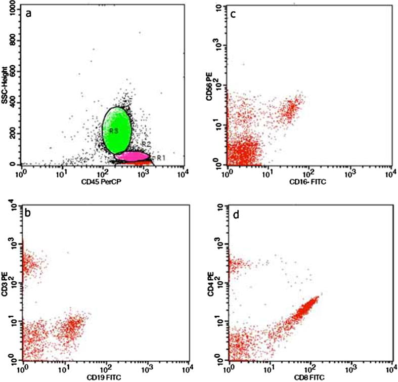
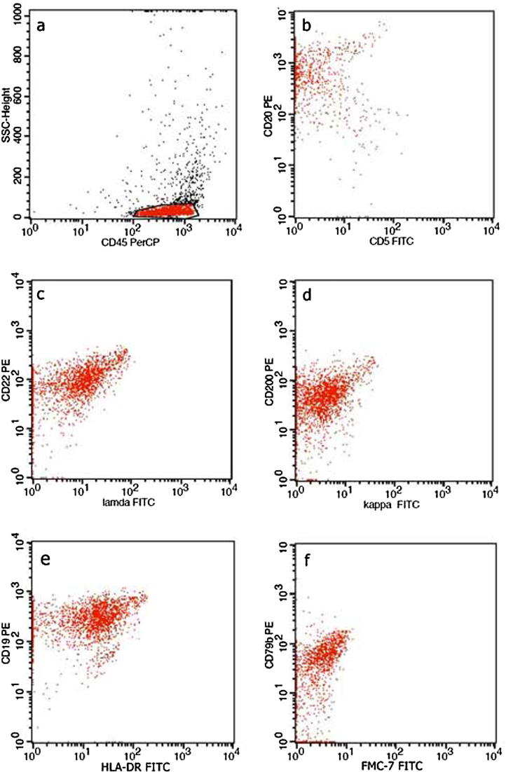
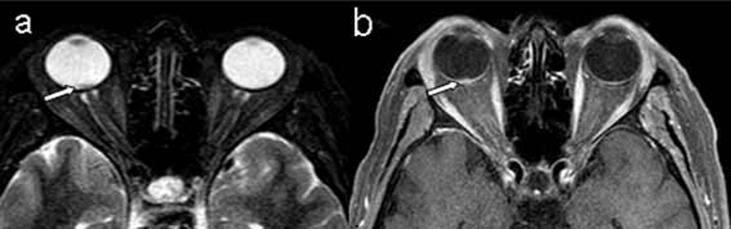
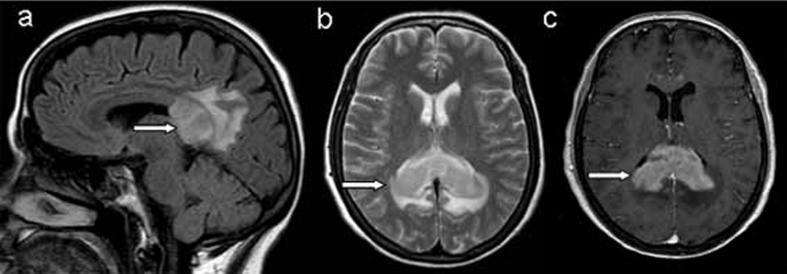
Primary vitreoretinal lymphoma (PVRL) is a rare ocular lymphoid malignancy, which consists a subset of primary central system lymphoma (PCNSL) and the most common type of intraocular lymphoma. The involvement of eyes is estimated to be approximately 20% of PCNSL, but the brain involvement may be up to 80% of PVRL. Typically, PVRL is a high grade B-cell malignancy of the retina and needs to be assorted from choroidal low-grade B-cell lymphomas. Very often PVRL masquerades and can be erroneously diagnosed as chronic uveitis, white dot syndromes or other neoplasms. Establishing an accurate diagnosis may involve cytology/pathology, immunohistochemistry, flow cytometry, molecular pathology and cytokine profile analysis. There is inadequate information about PVRL's true incidence, ethnic/geographical variation and pathogenetic mechanisms. The therapeutic approach of PVRL involves aggressive chemotherapy and radiation therapy. Although PVRL tends to have a good response to the initial treatment, the prognosis is poor and the survival restricted due to the high relapse rates and CNS involvement.

Keywords: B-cell lymphoma; Intraocular lymphoma; Masquerade syndrome; Primary CNS lymphoma; Primary vitreoretinal lymphoma.


Figures

Fig. 1
Fig. 1
(a) Fundus image with optic disc edema and obscured details due to vitreous haze in a 65 year-old lady (affected right eye), (b) SD-OCT: papillomacular bundle edema, nodular hyper-reflective infiltrations on the level of retinal pigment epithelium (RPE) along with partial destruction of RPE, c) B-mode of the same eye: moderately severe vitritis.
Fig. 2
Fig. 2
(a) Fluorescein angiography: optic disc edema and hyperfluorescent dots in the posterior pole, (b) Signs of retinal vasculitis (hemorrhages) and optic disc edema extended mainly to the nasal area of the posterior pole, (c) SD-OCT: increased papillomacular bundle edema compared to the initially documented (Fig. 1b) (d) B-mode: remnants of vitritis.
Fig. 3
Fig. 3
(a) SD-OCT 1 week before the end of the treatment: significant decrease of papillomacular bundle edema, along with reduction of number of hyper-reflective infiltrations. (b) Clouding of fundus details (VA: counting fingers at 0.5 m) due to recurrence of vitritis, with a deterioration of optic disc edema and signs of vasculitis. Yellow retinal infiltrates are increased in number.
Fig. 4
Fig. 4
(a) Lymphoid cells of medium size admixed with histiocytes. (Thin Prep smear, Papanicolaou stain; X 600), (b) Lymphoid cells with atypical morphologic features. (Thin Prep smear, Papanicolaou stain; X 600).
Fig. 5
Fig. 5
(a) Characteristic dot plots of CDs expression on peripheral blood lymphocytes subpopulations T- lymphocytes (CD3+) (b), B-lymphocytes (CD19+) (b), NK cells (CD16+56 positive) (c), T -helper cells (CD4+) and T -cytotoxic cells (CD8+) (d). Fig. 5b. (a) Characteristic dot plots of CDs expression on vitreous aspirations, showing the characteristic phenotypic profile CD20+ CD5- (b), CD22+ CD λ dim+ (c), CD200+, CDK- (d), CD19+ HLADR+ (e), FMC7- CD79b+ (f). The above findings consist of B lymphoma cells.
Fig. 5
Fig. 5
(a) Characteristic dot plots of CDs expression on peripheral blood lymphocytes subpopulations T- lymphocytes (CD3+) (b), B-lymphocytes (CD19+) (b), NK cells (CD16+56 positive) (c), T -helper cells (CD4+) and T -cytotoxic cells (CD8+) (d). Fig. 5b. (a) Characteristic dot plots of CDs expression on vitreous aspirations, showing the characteristic phenotypic profile CD20+ CD5- (b), CD22+ CD λ dim+ (c), CD200+, CDK- (d), CD19+ HLADR+ (e), FMC7- CD79b+ (f). The above findings consist of B lymphoma cells.
Fig. 6
Fig. 6
(a) STIR-weighted transverse image, (b) T1-weighted transverse image after contrast injection reveal a small enhanced mass lesion in the dorsal portion of the right eye ball (arrows).
Fig. 7
Fig. 7
(a) Sagittal T2-weighted FLAIR image in the midline shows an extensive mass lesion in splenium of the corpus callosum, (b) Axial T2-weighted image reveals an infiltrating mass lesion without evidence of necrosis, (c) Axial T1-weighted image after contrast injection (Gd-DTPA 0.1 mmol/kg) shows intensely and diffusely enhancement of the lesion (arrows). The magnetic resonance imaging findings are compatible with lymphoma.

References

    1. Coupland S.E., Damato B. Understanding intraocular lymphomas. Clin Experiment Ophthalmol. 2008;36:564–578. - PubMed
    1. Chan C.C., Rubenstein J.L., Coupland S.E. Primary vitreoretinal lymphoma: a report from an International Primary Central Nervous System Lymphoma Collaborative Group symposium. Oncologist. 2011;16:1589–1599. - PMC - PubMed
    1. Sagoo M.S., Mehta H., Swampillai A.J. Primary intraocular lymphoma. Surv Ophthalmol. 2014;59:503–516. - PubMed
    1. Aziz H.A., Peereboom D.M., Singh A.D. Primary central nervous system lymphoma. Int Ophthalmol Clin. 2015;55:111–121. - PubMed
    1. Fend F., Ferreri A.J., Coupland S.E. How we diagnose and treat vitreoretinal lymphoma. Br J Haematol. 2016;173:680–692. - PubMed

LinkOut - more resources