. 2019 Mar 15:13:101.
doi: 10.3389/fncel.2019.00101. eCollection 2019.

Homer1a Attenuates Endoplasmic Reticulum Stress-Induced Mitochondrial Stress After Ischemic Reperfusion Injury by Inhibiting the PERK Pathway

Affiliations

Homer1a Attenuates Endoplasmic Reticulum Stress-Induced Mitochondrial Stress After Ischemic Reperfusion Injury by Inhibiting the PERK Pathway

Jialiang Wei et al. Front Cell Neurosci. .

Abstract

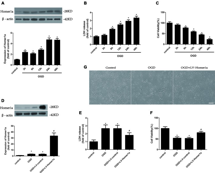
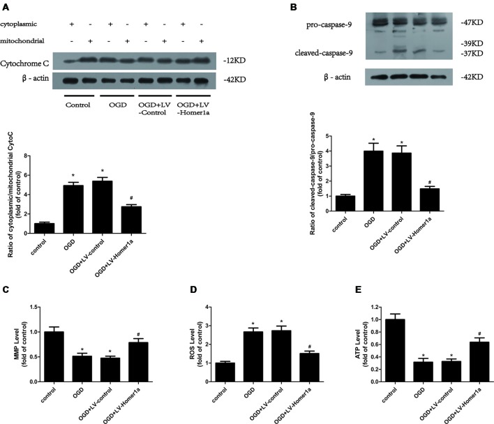
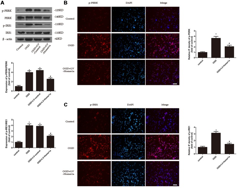
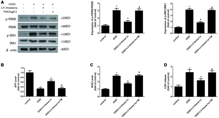
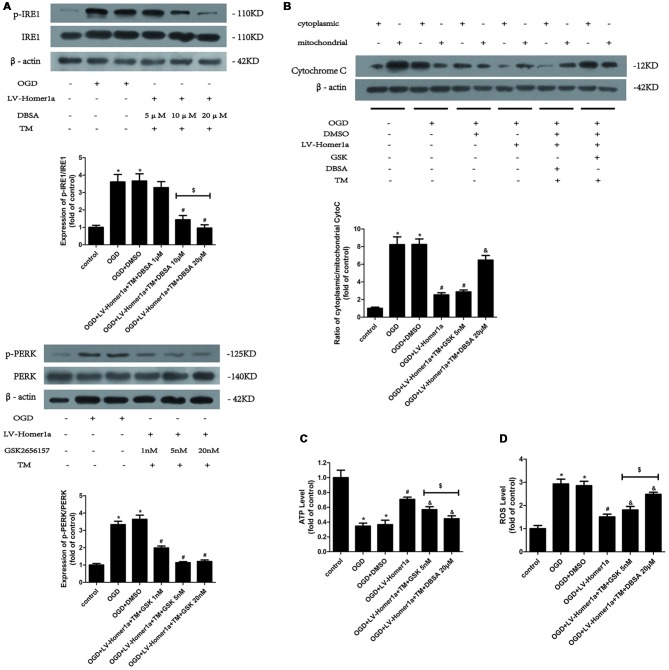
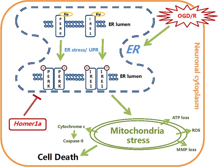
Homer1a is the short form of a scaffold protein that plays a protective role in many forms of stress. However, the role of Homer1a in cerebral ischemia/reperfusion (I/R) injury and its potential mechanism is still unknown. In this study, we found that Homer1a was upregulated by oxygen and glucose deprivation (OGD) and that overexpression of Homer1a alleviated OGD-induced lactate dehydrogenase (LDH) release and cell death in cultured cortical neurons. After OGD treatment, the overexpression of Homer1a preserved mitochondrial function, as evidenced by less cytochrome c release, less reactive oxygen species (ROS) production, less ATP and mitochondrial membrane potential (MMP) loss, less caspase-9 activation, and inhibition of endoplasmic reticulum (ER) stress confirmed by the decreased expression of phosphate-PKR-like ER Kinase (p-PERK)/PERK and phosphate- inositol-requiring enzyme 1 (p-IRE1)/IRE1 and immunofluorescence (IF) staining. In addition, mitochondrial protection of Homer1a was blocked by the ER stress activator Tunicamycin (TM) with a re-escalated ROS level, increasing ATP and MMP loss. Furthermore, Homer1a overexpression-induced mitochondrial stress attenuation was significantly reversed by activating the PERK pathway with TM and p-IRE1 inhibitor 3,5-dibromosalicylaldehyde (DBSA), as evidenced by increased cytochrome c release, increased ATP loss and a higher ROS level. However, activating the IRE1 pathway with TM and p-PERK inhibitor GSK2656157 showed little change in cytochrome c release and exhibited a moderate upgrade of ATP loss and ROS production in neurons. In summary, these findings demonstrated that Homer1a protects against OGD-induced injury by preserving mitochondrial function through inhibiting the PERK pathway. Our finding may reveal a promising target of protecting neurons from cerebral I/R injury.

Keywords: PERK kinase; endoplasmic reticulum stress; homer1a; ischemic stroke; mitochondrial dysfunction.


Figures

Figure 1
Figure 1
Homer1a protects neurons against ischemia/reperfusion (I/R) injury. Cultured cortical neurons were treated with oxygen and glucose deprivation (OGD) for 1h, and the expression level of Homer1a protein at different reperfusion time points were investigated by Western blot (A). Then the cytotoxicity of neurons was detected by lactate dehydrogenase (LDH) release assay (B) and the cell viability was evaluated by CCK-8 test (C). The cortical neurons were then transfected with LV-Homer1a or LV-control for 72 h and exposed to OGD. The Homer1a protein expression was detect by Western blot (D), and cytotoxicity and cell viability were tested in each group (E,F). The cortical neurons were also observed under representative phase photomicrographs (G). The data are represented as means ± SEM. *p < 0.05 vs. Control, #p < 0.05 vs. OGD.
Figure 2
Figure 2
Homer1a conserves mitochondrial function in neurons after I/R injury. Cultured cortical neurons were transfected with LV-Homer1a or LV-control for 72 h and exposed to OGD. The cytochrome c release to cytoplasm and the activation of caspase-9 were detected by Western blot (A,B). The mitochondrial membrane potentials (MMPs) were detected by Rh123 test (C), the intracellular reactive oxygen species (ROS) level was investigated through H2-DCFDA (D) and the intracellular ATP level was measured by ATP assay (E). The data are represented as means ± SEM. *p < 0.05 vs. Control, #p < 0.05 vs. OGD.
Figure 3
Figure 3
Homer1a reduces endoplasmic reticulum (ER) stress in neurons that underwent I/R injury. Cultured cortical neurons were transfected with LV-Homer1a or LV-control for 72 h and exposed to OGD. Levels of phosphorylated-PKR-like ER kinase (p-PERK), PERK, phosphorylated-IRE1 (p-IRE1) and IRE1 were measured with Western blot (A). The level of p-PERK and p-IRE1 were also determined with immunofluorescence (IF) staining. Images were captured with fluorescence microscope. Red: p-PERK or p-IRE1, blue: nuclei-staining by DAPI. Scale bar = 50 μm. The IF intensity of each group was calculated with ImageJ (B,C). The data are represented as means ± SEM. *p < 0.05 vs. Control, #p < 0.05 vs. OGD.
Figure 4
Figure 4
Homer1a alleviates mitochondria dysfunction by reducing ER stress after I/R injury. Cultured cortical neurons were pre-treated with 2 μg/L Tunicamycin (TM) 24 h to activate the ER stress and exposed to I/R injury. The effects of TM were tested by Western blot (A). The intracellular ATP level was measured by ATP assay (B) and the intracellular ROS level was investigated through H2-DCFDA (C). We also investigated the neurons cytotoxicity of each group with LDH release assay (D). The data are represented as means ± SEM. *p < 0.05 vs. Control, #p < 0.05 vs. OGD, &p < 0.05 vs. OGD+LV-Homer1a.
Figure 5
Figure 5
Homer1a preserves mitochondrial function by reducing ER stress mainly via the PERK pathway after I/R injury. TM was used to activate the ER stress whereas the GSK2656157 and 3,5-dibromosalicylaldehyde (DBSA) were used to inhibit the PERK and IRE1 respectively. Different DBSA concentrations were tested in cortical neurons and detected by Western blot (A) as well as GSK2656157 (B). Then 20 μM DBSA and 5 nM GSK2656157 were chosen to apply to follow experiments. The cytochrome c release to cytoplasm in each group was detected through calculating the ratio of cytoplasmic/mitochondrial cytochrome c by Western blot (B). The intracellular ATP level was measured by ATP assay (C) and the intracellular ROS level was investigated through H2-DCFDA (D). The data are represented as means ± SEM. $p < 0.05, *p < 0.05 vs. Control, #p < 0.05 vs. OGD, &p < 0.05 vs. OGD+LV-Homer1a.
Figure 6
Figure 6
The schematic diagram depicts the molecular mechanisms of homer1a-induced neuron-protection against OGD injury. Homer1a overexpression inhibited the OGD-induced activation of ER stress, which alleviated the secondary mitochondria dysfunction by regulating ATP, MMP, ROS, cytochrome c and caspase-9. In addition, Homer1a-induced preservation of mitochondrial function was achieved mainly via inhibiting the PERK pathway.

References

    1. Benjamin E. J., Virani S. S., Callaway C. W., Chamberlain A. M., Chang A. R., Cheng S., et al. . (2018). Heart Disease and stroke statistics-2018 update: a report from the american heart association. Circulation 137, e67–e492. 10.1161/CIR.0000000000000558 - DOI - PubMed
    1. Bennetts P. S., Pierce J. D. (2010). Apoptosis: understanding programmed cell death for the CRNA. AANA J. 78, 237–245. - PubMed
    1. Bhatti J. S., Bhatti G. K., Reddy P. H. (2017). Mitochondrial dysfunction and oxidative stress in metabolic disorders—a step towards mitochondria based therapeutic strategies. Biochim. Biophys. Acta Mol. Basis Dis. 1863, 1066–1077. 10.1016/j.bbadis.2016.11.010 - DOI - PMC - PubMed
    1. Bravo-Sagua R., Rodriguez A. E., Kuzmicic J., Gutierrez T., Lopez-Crisosto C., Quiroga C., et al. . (2013). Cell death and survival through the endoplasmic reticulum-mitochondrial axis. Curr. Mol. Med. 13, 317–329. 10.2174/156652413804810781 - DOI - PMC - PubMed
    1. Chen T., Fei F., Jiang X. F., Zhang L., Qu Y., Huo K., et al. . (2012). Down-regulation of Homer1b/c attenuates glutamate-mediated excitotoxicity through endoplasmic reticulum and mitochondria pathways in rat cortical neurons. Free Radic. Biol. Med. 52, 208–217. 10.1016/j.freeradbiomed.2011.10.451 - DOI - PubMed

LinkOut - more resources