Review
. 2019 Mar 15:7:71.
doi: 10.3389/fped.2019.00071. eCollection 2019.

Epstein-Barr Virus-Associated T and NK-Cell Lymphoproliferative Diseases

Affiliations
Review

Epstein-Barr Virus-Associated T and NK-Cell Lymphoproliferative Diseases

Wook Youn Kim et al. Front Pediatr. .

Abstract

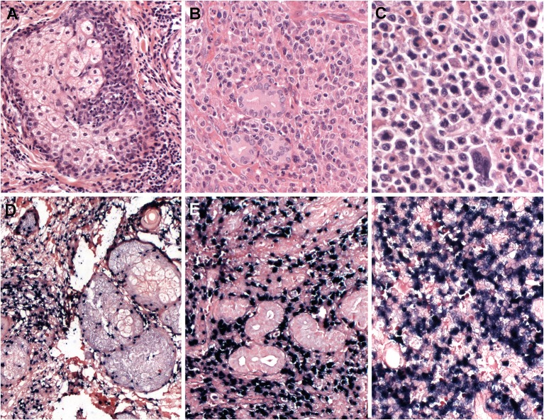
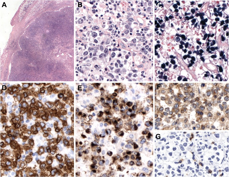
EBV-associated T and NK-cell lymphoproliferative diseases (EBV-T/NK LPDs) are characterized by the transformation and proliferation of EBV-infected T or NK cells. The 2016 revised World Health Organization classification recognizes the following EBV-positive lymphoproliferative disorders (LPD): chronic active EBV infection (CAEBV) of T- and NK-cell type (cutaneous and systemic forms), systemic EBV-positive T-cell lymphoma of childhood, aggressive NK-cell leukemia, extranodal NK/T-cell lymphoma, nasal type, and the new provisional entity primary EBV-positive nodal T/NK-cell lymphoma. EBV-associated hemophagocytic lymphohistiocytosis (HLH), although not included in the WHO classification because it is a reactive, inflammatory disease, is included in this review because it can be life-threatening and may have overlapping features with other EBV+ T/NK LPDs. EBV+ T/NK LPDs are rare diseases difficult to diagnose and manage properly, because some LPDs have unusual presentations, and discrepancies between clinical and histological findings might be encountered. Furthermore, EBV+ T/NK disorders share some clinico-pathological features, and may evolve into other categories during the clinical course, including malignant transformation of CAEBV. Here, we review the EBV+ T/NK LPDs in terms of their definitions, clinical features, histology, immunophenotype, molecular findings, and pathogenesis. This review aims to increase our understanding and awareness of the differential diagnosis among the different EBV+ T/NK LPDs. New insights into the genetic characteristics of these disorders will also be discussed.

Keywords: EBV; aggressive NK-cell leukemia; chronic active EBV infection; extranodal NK/T-cell lymphoma; hemophagocytic lymphohistiocytosis; lymphoproliferations; primary EBV nodal T and NK-cell lymphoma; systemic EBV positive T-cell lymphoma.


Figures

Figure 1
Figure 1
Hydroa vacciniforme-like lymphoproliferative disorder. (A) A skin biopsy with a subtle dermal infiltrate surrounding adnexae and blood vessels (H&E, 25x); (B) The lymphoid cells are EBV positive, as demonstrated by in situ hybridization for EBV-encoded small RNA (EBER) (in situ hybridization, 25x); (C) CD8 is positive in the majority of the infiltrating cells (immunohistochemistry, 25x); (D) The infiltrating cells are negative for CD4. CD4 highlights the abundant histiocytes (immunohistochemistry, 25x); (E) The infiltrating cells are predominantly small, without atypia (H&E, 400×); (F) The infiltrating cells are surrounding a blood vessel highlighted by CD8 stain (immunohistochemistry, 400×).
Figure 2
Figure 2
Systemic Epstein-Bar virus (EBV)-positive T-cell lymphoma of childhood. (A) Lymph node with partial preservation of the architecture with depleted germinal centers and expansion of the interfollicular area (H&E, 50×); (B) Many of the lymphoid cells in the interfollicular area are EBV-positive, as demonstrated by in situ hybridization for EBV-encoded small RNA (EBER) (in-situ hybridization 100×); (C) The neoplastic cells are mostly medium to large-sized cells with irregular nuclei. Note the presence of apoptosis (H&E, 400×); (D) Many cells are EBER positive (in-situ hybridization, 400×). LMP1 is positive indicating an EBV latency type 2 (immunohistochemistry, insert, 400×); (E) The neoplastic cells are CD8 positive (immunohistochemistry, 400×). Double stainings show that the CD8-positive cells (red) are EBER-positive (Black) (Immunohistochemistry and in situ hybridization, insert, 400×) (F) TIA1 is positive in the infiltrating cells (immunostaining, 400×).
Figure 3
Figure 3
Aggressive NK-cell leukemia. (A) The spleen shows a scant atypical lymphoid infiltrate of small cells with bland cytology surrounding blood vessels. Note the striking erythrophagocytosis (arrows) (H&E, 400×); (B) The liver shows an atypical infiltrate in the sinusoids composed of medium-sized cells with irregular nuclei and pale cytoplasm; (C) Neoplastic cells in the spleen are stained positively with EBER (in-situ hybridization 400×). Insert shows double staining of CD56 (red) and EBER (black) demonstrating that the NK cells are infected by EBV (Immunohistochemistry and in situ hybridization 400×); and (D) The infiltrating cells are CD56 positive (immunohistochemistry 400×); (E) The neoplastic cells in the liver are EBER positive. Note the intrasinusoidal infiltration characteristic of the disease (in-situ hybridization, 400×).
Figure 4
Figure 4
Extranodal, NK/T-cell lymphoma, nasal type in the skin. (A) Panoramic view of a skin biopsy shows a partially circumscribed nodule located in the subcutaneous tissue (H&E, scanned slide); (B) The tumor cells are CD56 positive (immunohistochemistry, scanned slide). (C) The lymphoid cells are positive for EBV-encoded small RNA in situ hybridization (EBER) (in situ hybridization) (D) The infiltrate is composed of large atypical cells with irregular nuclei. The tumor cells surround the adipocytes revealing a “lace-like pattern” mimicking panniculitis-like T-cell lymphoma. Numerous apoptotic bodies are observed (H&E, 400×); (E,F) Higher magnification demonstrates that the neoplastic cells are positive for CD56 and EBER, (immunohistochemistry and in situ hybridization 400×).
Figure 5
Figure 5
Extranodal, NK/T-cell lymphoma, nasal type. (A–C) Nasal biopsies displaying the morphological spectrum of ENKTCL. (A) Infiltrate of small lymphoid cells with bland cytology surrounding the sebaceous gland, mimicking a reactive lesion (H&E, 400×);(B) Dense lymphoid infiltrate of intermediate-sized cells, showing nuclear irregularity and pale to clear cytoplasm (H&E, 400×); (C) Atypical cell infiltrate, of pleomorphic large cells admixed with intermediate-sized cells (H&E, 400×); (D–F) Lymphoma cells are positive for EBV-encoded small RNA in situ hybridization (EBER, 400×).
Figure 6
Figure 6
Primary EBV-positive nodal T and NK-cell lymphoma. (A) Lymph node with complete effacement of the architecture by a diffuse infiltrate that extends beyond the capsule and infiltrate the perinodal fat (H&E, 12,5×); (B) Neoplastic cells are large, pleomorphic with irregular nuclei and clear or pale cytoplasm (HE,400×); (C–E) The neoplastic cells EBER, CD56 and TIA-1 positive (in-situ hybridization and immunohistochemistry, 400×); (F) TCR-gamma immunostain demonstrates the gamma-delta derivation of the tumor cells (immunohistochemistry, 400×); (G) TCR alpha-beta (BetaF1) is negative in the tumor cells but positive in the reactive T cells (immunohistochemistry, 400×).

References

    1. Young LS, Rickinson AB. Epstein-Barr virus: 40 years on. Nat Rev Cancer. (2004) 4:757–68. 10.1038/nrc1452 - DOI - PubMed
    1. Taylor GS, Long HM, Brooks JM, Rickinson AB, Hislop AD. The immunology of Epstein-Barr virus-induced disease. Annu Rev Immunol. (2015) 33:787–821. 10.1146/annurev-immunol-032414-112326 - DOI - PubMed
    1. Quintanilla-Martinez L, Ko Y-H, Kimura H, Jaffe ES. EBV-positive T-cell and NK-cell lymphoproliferative diseases of childhood. In: Swerdlow SH, Campo E, Harris NL, Jaffe ES, Pileri SA, Stein H, et al. editors. Who Classification of Tumours of Haematopoietic and Lymphoid Tissues. Lyon, France: IARC; (2017). p. 355–63.
    1. Kimura H, Ito Y, Kawabe S, Gotoh K, Takahashi Y, Kojima S, et al. EBV-associated T/NK-cell lymphoproliferative diseases in nonimmunocompromised hosts: prospective analysis of 108 cases. Blood. (2012) 119:673–86. 10.1182/blood-2011-10-381921 - DOI - PubMed
    1. Ishii E, Ohga S, Imashuku S, Yasukawa M, Tsuda H, Miura I, et al. Nationwide survey of hemophagocytic lymphohistiocytosis in Japan. Int J Hematol. (2007) 86:58–65. 10.1532/IJH97.07012 - DOI - PubMed

LinkOut - more resources