

PRGF as adjunct to DBB in maxillary sinus floor augmentation: histological results of a pilot split-mouth study
- PMID: 30931490
- PMCID: PMC6441666
- DOI: 10.1186/s40729-019-0166-6
PRGF as adjunct to DBB in maxillary sinus floor augmentation: histological results of a pilot split-mouth study
Abstract
Background: Various technologies of autologous blood concentrates are currently evaluated for their potential to enhance bone formation.
Aim: To report on the histological outcome of maxillary sinus floor augmentation (MSFA) with deproteinized bovine bone (DBB) in combination with chair-side prepared autologous platelet-rich growth factor (PRGF), in comparison to that with DBB alone.
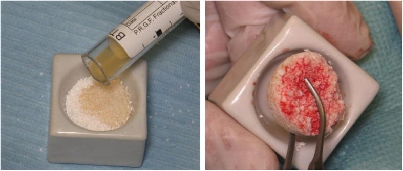
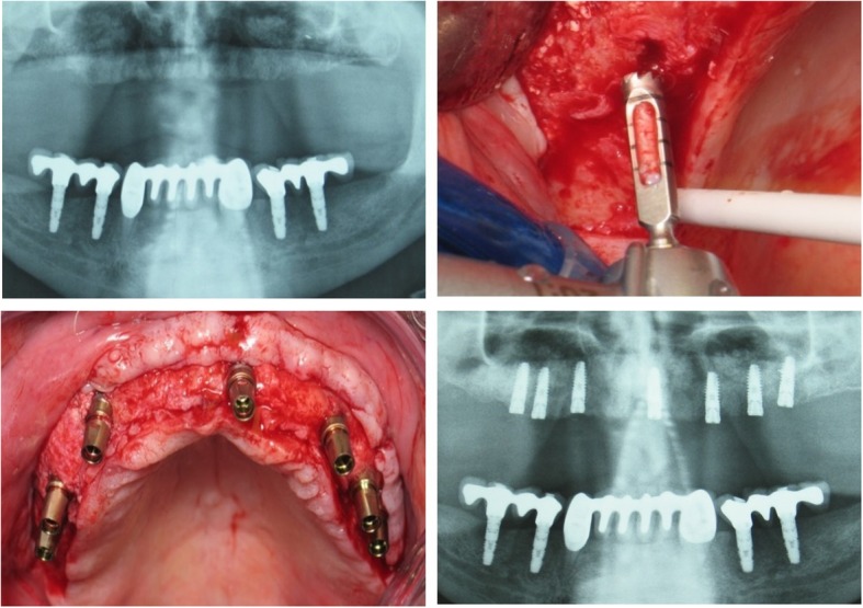
Materials and methods: Six partially edentulous patients with ≤ 3-mm residual bone height bilaterally in the posterior maxilla were subjected to MSFA with the lateral window technique, using DBB in combination with PRGF (PRGF System1 Vitoria, Spain) on one side or DBB alone on the contralateral side. Cylindrical biopsies from the augmented sinuses were collected during implant installation, ca. 6 months post-MSFA, and subjected to non-decalcified histological and histomorphometric evaluation.
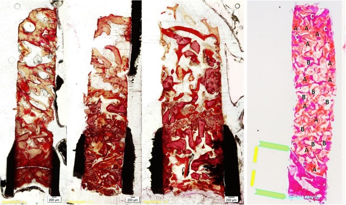
Results: The collected biopsies varied in length (range 3.5-9.9 mm); consequently, the portion of the biopsy representing augmented tissues also varied (range 2.3-14.6 mm2). New bone formation with a trabecular appearance and numerous DBB particles in contact with the new bone or with loose connective tissue were observed. No differences in the relative volumes of bone formation were found in sinuses augmented with DBB + PRGF or DBB alone 6 months after MSFA (35.6 ± 8.26 mm and 37.8 ± 3.15 mm, respectively).
Conclusion and clinical implications: In conclusion, based on these preliminary results, PRGF as adjunct to DBB for MSFA, except from improved handling during the operation, does not appear to enhance nor interfere with bone formation inside the human sinus 6 months after MSFA, compared with the use of DBB alone.
Keywords: Deproteinized bovine bone; Histology; Humans; PRGF; Sinus elevation.
Conflict of interest statement
Ethics approval and consent to participate
Patient recruitment and data collection of this study took place at the Aristotle University of Thessaloniki, School of Dentistry, Greece. The research was approved by the Ethics Committee of the Aristotle University of Thessaloniki, Greece (14/02-02-2017) and all activities were conducted in full accordance with ethical principles, including the World Medical Association Declaration of Helsinki. We described the purpose of the study to patients, and the data were obtained anonymously.
Consent for publication
Not applicable.
Competing interests
Authors Batas Leonidas, Tsalikis Lazaros, and Stavropoulos Andreas declare that they have no competing interests.
Publisher’s Note
Springer Nature remains neutral with regard to jurisdictional claims in published maps and institutional affiliations.
Figures

Similar articles
-
Clinical and radiographic outcomes of dental implant after maxillary sinus floor augmentation with rhBMP-2/hydroxyapatite compared to deproteinized bovine bone.PLoS One. 2022 Aug 25;17(8):e0273399. doi: 10.1371/journal.pone.0273399. eCollection 2022. PLoS One. 2022. PMID: 36007001 Free PMC article.
-
Deproteinized bovine bone and freeze-dried bone allograft in sinus floor augmentation: A randomized controlled trial.Clin Implant Dent Relat Res. 2023 Apr;25(2):343-351. doi: 10.1111/cid.13179. Epub 2023 Jan 11. Clin Implant Dent Relat Res. 2023. PMID: 36628938 Clinical Trial.
-
Influence of Healing Period Upon Bone Turn Over on Maxillary Sinus Floor Augmentation Grafted Solely with Deproteinized Bovine Bone Mineral: A Prospective Human Histological and Clinical Trial.Clin Implant Dent Relat Res. 2017 Apr;19(2):341-350. doi: 10.1111/cid.12463. Epub 2016 Nov 16. Clin Implant Dent Relat Res. 2017. PMID: 27862924 Clinical Trial.
-
Long-term effectiveness of maxillary sinus floor augmentation: A systematic review and meta-analysis.J Clin Periodontol. 2019 Jun;46 Suppl 21:307-318. doi: 10.1111/jcpe.13055. J Clin Periodontol. 2019. PMID: 30624789
-
Plasma rich in growth factors (PRGF) in intraoral bone grafting procedures: A systematic review.J Craniomaxillofac Surg. 2019 Mar;47(3):443-453. doi: 10.1016/j.jcms.2019.01.012. Epub 2019 Jan 17. J Craniomaxillofac Surg. 2019. PMID: 30711470
Cited by
-
Residual Bone Height and New Bone Formation after Maxillary Sinus Augmentation Procedure Using Biomaterials: A Network Meta-Analysis of Clinical Trials.Materials (Basel). 2023 Feb 6;16(4):1376. doi: 10.3390/ma16041376. Materials (Basel). 2023. PMID: 36837005 Free PMC article. Review.
-
Clinical Indications and Outcomes of Sinus Floor Augmentation With Bone Substitutes: An Evidence-Based Review.Clin Implant Dent Relat Res. 2025 Feb;27(1):e13400. doi: 10.1111/cid.13400. Epub 2024 Oct 17. Clin Implant Dent Relat Res. 2025. PMID: 39415739 Free PMC article. Review. No abstract available.
-
Relative performance of various biomaterials used for maxillary sinus augmentation: A Bayesian network meta-analysis.Clin Oral Implants Res. 2021 Feb;32(2):135-153. doi: 10.1111/clr.13690. Epub 2021 Jan 6. Clin Oral Implants Res. 2021. PMID: 33230862 Free PMC article. Review.
-
A retrospective preliminary histomorphometric and clinical investigation on sinus augmentation using enzyme-deantigenic, collagen-preserving equine bone granules and plasma rich in growth factors.Int J Implant Dent. 2021 Jun 11;7(1):60. doi: 10.1186/s40729-021-00336-9. Int J Implant Dent. 2021. PMID: 34114113 Free PMC article.
-
Evaluation of Bone Gain and Complication Rates after Guided Bone Regeneration with Titanium Foils: A Systematic Review.Materials (Basel). 2020 Nov 25;13(23):5346. doi: 10.3390/ma13235346. Materials (Basel). 2020. PMID: 33255825 Free PMC article. Review.
References
-
- Boyne PJ, James RA. Grafting of the maxillary sinus floor with autogenous marrow and bone. J Oral Surg. 1980;38(8):613–616. - PubMed
-
- Summers RB. The osteotome technique: Part 3--Less invasive methods of elevating the sinus floor. Compendium. 1994;15(6):698–700. - PubMed
-
- Toffler M. Site development in the posterior maxilla using osteocompression and apical alveolar displacement. Compend Contin Educ Dent. 2001;22(9):775–780. - PubMed
-
- Del Fabbro M, et al. Systematic review of survival rates for implants placed in the grafted maxillary sinus. Int J Periodontics Restorative Dent. 2004;24(6):565–577. - PubMed
-
- Shanbhag S, Shanbhag V, Stavropoulos A. Volume changes of maxillary sinus augmentations over time: a systematic review. Int J Oral Maxillofac Implants. 2014;29(4):881–92. - PubMed
LinkOut - more resources
Full Text Sources
