. 2019 Apr 1;60(5):1328-1335.
doi: 10.1167/iovs.18-25883.

Low-Intensity Exercise in Mice Is Sufficient to Protect Retinal Function During Light-Induced Retinal Degeneration

Affiliations

Low-Intensity Exercise in Mice Is Sufficient to Protect Retinal Function During Light-Induced Retinal Degeneration

Lukas M Mees et al. Invest Ophthalmol Vis Sci. .

Abstract

Purpose: We previously reported that a specific treadmill running exercise regimen protects against light-induced retinal degeneration (LIRD) in mice. We hypothesized that this protective effect varies with running intensity. To test this, mice undergoing LIRD were run at different treadmill speeds and retinal function was assessed.

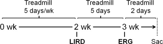
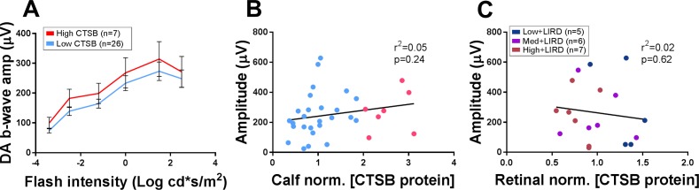
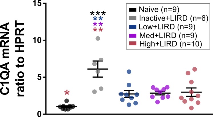
Methods: BALB/c mice were assigned to LIRD groups at varying treadmill speeds-0, 5, 10, or 20 m/min labeled inactive, low, medium, and high, respectively-and compared with naïve mice exposed to standard lighting (50 lux; naïve). Following 2 weeks of exercise, a subset of mice were exposed to toxic light (10,000 lux; LIRD) for 4 hours. After 5 additional days of exercise, retinal function was assessed by ERG. Corticosterone levels in serum and cathepsin B (CTSB) protein levels in muscle, brain, serum, and retina were measured. The retinal gene expression of complement factor 1qa (C1qa) and CTSB were measured.

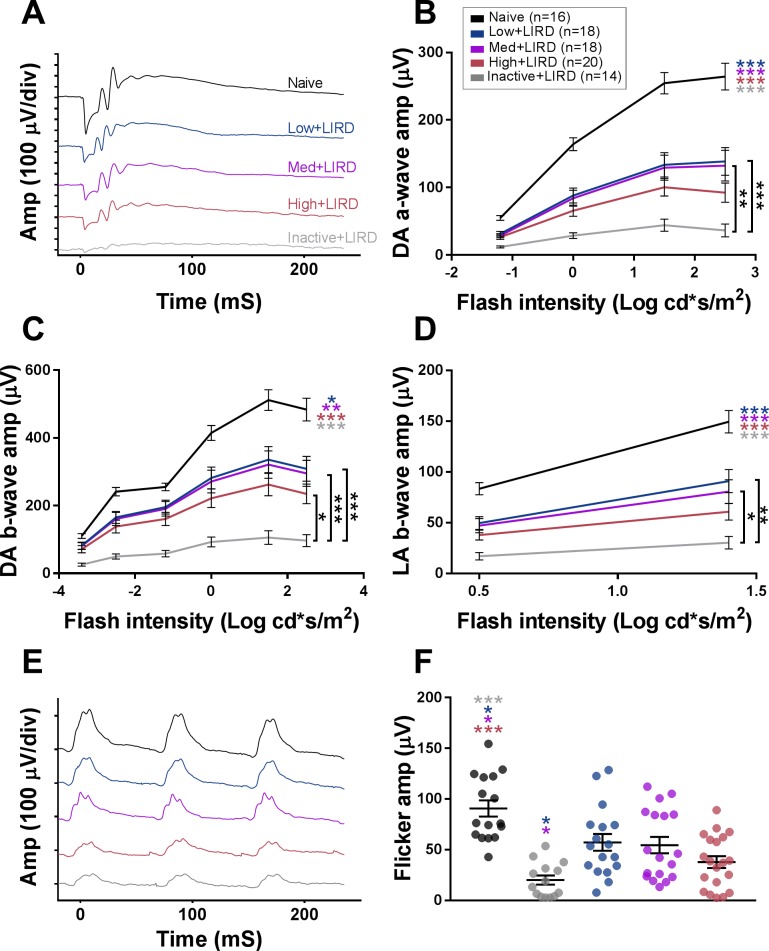
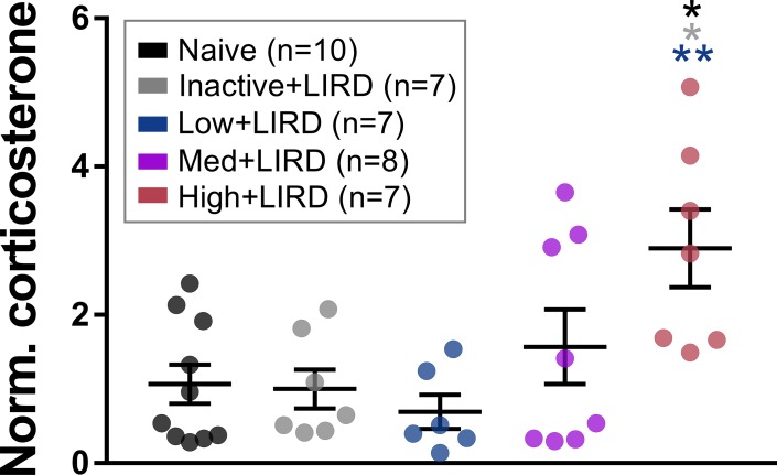
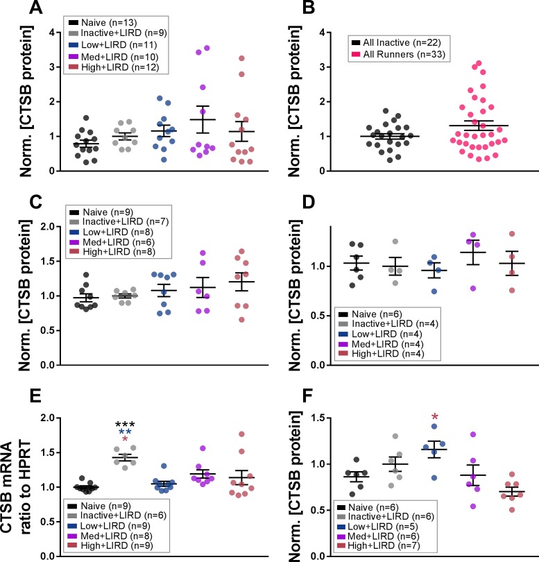
Results: The low+LIRD and medium+LIRD exercise groups had greater a- and b-wave ERG amplitudes when compared with the inactive+LIRD group (P < 0.02). The high+LIRD mice only differed from the inactive+LIRD mice in their dark-adapted b-waves. Serum corticosterone increased in the high+LIRD mice (P < 0.006). Retinal CTSB protein levels were higher in the low+LIRD versus high+LIRD mice (P < 0.004) but were otherwise unchanged. Exercise of any intensity decreased C1qa gene expression.

Conclusions: Faster running did not additionally protect against LIRD, but it did increase serum corticosterone, suggesting stress-induced limits to exercise benefits. Unexpectedly, exercise did not increase CTSB proteins levels in muscle or serum, suggesting that it may not mediate exercise effects. Our results have implications for the use of low-intensity exercise as a vision loss treatment.


Figures

Figure 1
Figure 1
Experimental design. Timeline shown in the middle. Running procedure on top, and experimental procedures on bottom. Wk, week; sac, sacrifice.
Figure 2
Figure 2
Effect of exercise intensity on retinal function. Representative ERG waveforms are shown for maximum dark-adapted (DA) stimuli (A, 2.5 log cd s/m2) and flicker (E, 2.0 log cd s/m2). Quantified ERG amplitudes are depicted for dark-adapted a-wave (B), dark-adapted b-wave (C), light-adapted (LA) b-wave (D), and flicker (F). Data are depicted as mean ± SEM. Circles on (F) represent measurements for individual animals. Colored asterisks refer to the difference between the group they are closest to and the group of the referenced color. Amp, amplitude, div, division. *P < 0.026, **P < 0.005, ***P < 0.001.
Figure 3
Figure 3
High-intensity exercise increases serum corticosterone. Serum corticosterone was elevated in mice exercised at high intensity, but not low or medium intensity. Data are depicted as mean ± SEM. Circles represent measurements for individual animals. Colored asterisks refer to the difference between the group they are closest to and the group of the referenced color. *P < 0.05, **P < 0.01.
Figure 4
Figure 4
CTSB gene expression and protein levels altered in retina after exercise. CTSB protein in calf muscle was analyzed by exercise level (A) and when comparing all inactive (inactive+LIRD and naïve) mice to all exercised mice (B). CTSB protein levels were also examined in the brain (C) and serum (D). In the retina, CTSB gene expression (E) and protein levels (F) were measured. Data are depicted as mean ± SEM normalized to inactive+LIRD for A and C–E and to all inactive in B. Circles represent measurements for individual animals. Colored asterisks refer to the difference between the group they are closest to and the group of the referenced color. *P < 0.05, **P < 0.01, ***P < 0.001.
Figure 5
Figure 5
No correlation in calf muscle or retinal CTSB to functional protection. (A) Active animals across all three exercise intensity groups were divided into high- and low-muscular CTSB levels. Animals with a calf muscle CTSB level 10% higher than the max inactive+LIRD animal were classified as “high CTSB” and compared with those below the threshold “low CTSB” for dark-adapted b-wave amplitude. The brightest flash dark-adapted b-wave amplitudes showed no correlation with calf muscle CTSB (B) and retinal CTSB protein (C). Circles represent measurements for individual animals. Data in A depicted as mean ± SEM. Linear regression line shown in B and C. Amp, amplitude.
Figure 6
Figure 6
Any exercise intensity lowers retinal C1qa gene expression. Retinal C1qa mRNA was elevated in inactive+LIRD animals, but preserved at naïve levels in mice exercised at any of the three treadmill speeds. Data are depicted as mean ± SEM. Circles represent measurements for individual animals. Colored asterisks refer to the difference between the group they are closest to and the group of the referenced color. *P < 0.05, **P < 0.01, ***P < 0.001.

References

    1. Radahmadi M, Alaei H, Sharifi MR, Hosseini N. Effect of forced exercise and exercise withdrawal on memory, serum and hippocampal corticosterone levels in rats. Exp Brain Res. 2015;233:2789–2799. - PubMed
    1. Cetinkaya C, Sisman AR, Kiray M, et al. Positive effects of aerobic exercise on learning and memory functioning, which correlate with hippocampal IGF-1 increase in adolescent rats. Neurosci Lett. 2013;549:177–181. - PubMed
    1. Nokia MS, Lensu S, Ahtiainen JP, et al. Physical exercise increases adult hippocampal neurogenesis in male rats provided it is aerobic and sustained. J Physiol. 2016;594:1855–1873. - PMC - PubMed
    1. Roig M, Nordbrandt S, Geertsen SS, Nielsen JB. The effects of cardiovascular exercise on human memory: a review with meta-analysis. Neurosci Biobehav Rev. 2013;37:1645–1666. - PubMed
    1. Cassilhas RC, Tufik S, de Mello MT. Physical exercise, neuroplasticity, spatial learning and memory. Cell Mol Life Sci. 2016;73:975–983. - PMC - PubMed

Publication types