

Intrafraction tumor motion during deep inspiration breath hold pancreatic cancer treatment
- PMID: 30933428
- PMCID: PMC6523018
- DOI: 10.1002/acm2.12577
Intrafraction tumor motion during deep inspiration breath hold pancreatic cancer treatment
Abstract
Purpose: Beam gating with deep inspiration breath hold (DIBH) has been widely used for motion management in radiotherapy. Normally it relies on some external surrogate for estimating the internal target motion, while the exact internal motion is unknown. In this study, we used the intrafraction motion review (IMR) application to directly track an internal target and characterized the residual motion during DIBH treatment for pancreatic cancer patients through their full treatment courses.
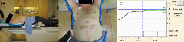
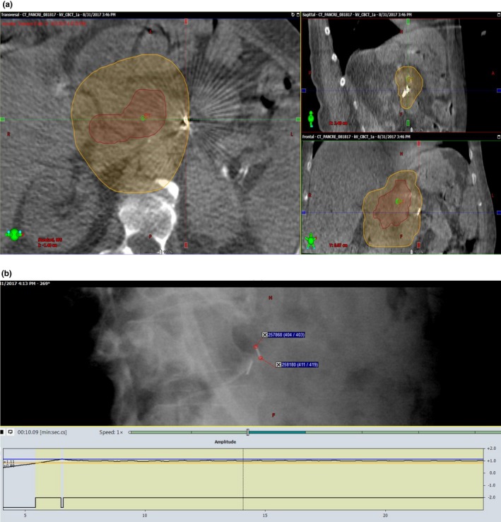
Methods and materials: Eight patients with pancreatic cancer treated with DIBH volumetric modulated arc therapy in 2017 and 2018 were selected for this study, each with some radiopaque markers (fiducial or surgical clips) implanted near or inside the target. The Varian Real-time Position Management (RPM) system was used to monitor the breath hold, represented by the anterior-posterior displacement of an external surrogate, namely reflective markers mounted on a plastic block placed on the patient's abdomen. Before each treatment, a cone beam computed tomography (CBCT) scan under DIBH was acquired for patient setup. For scan and treatment, the breath hold reported by RPM had to lie within a 3 mm window. IMR kV images were taken every 20° or 40° gantry rotation during dose delivery, resulting in over 5000 images for the cohort. The internal markers were manually identified in the IMR images. The residual motion amplitudes of the markers as well as the displacement from their initial positions located in the setup CBCT images were analyzed.
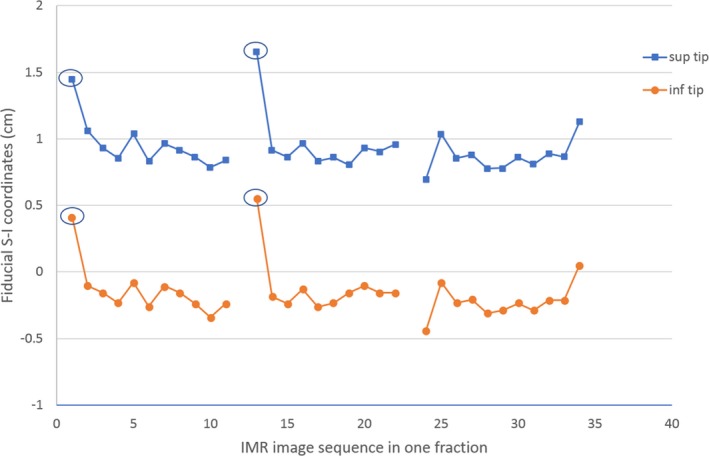
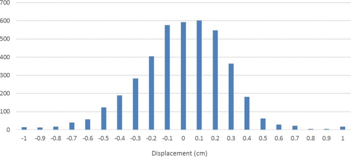
Results: Even though the external markers indicated that the respiratory motion was within 3 mm in DIBH treatment, significant residual internal target motion was observed for some patients. The range of average motion was from 3.4 to 7.9 mm, with standard deviation ranging from 1.2 to 3.5 mm. For all patients, the target residual motions seemed to be random with mean positions around their initial setup positions. Therefore, the absolute target displacement relative to the initial position was small during DIBH treatment, with the mean and the standard deviation 0.6 and 2.9 mm, respectively.
Conclusions: Internal target motion may differ from external surrogate motion in DIBH treatment. Radiographic verification of target position at the beginning and during each fraction is necessary for precise RT delivery. IMR can serve as a useful tool to directly monitor the internal target motion.
Keywords: deep inspiration breath hold; intrafraction motion; pancreatic cancer; radiation therapy.
© 2019 The Authors. Journal of Applied Clinical Medical Physics published by Wiley Periodicals, Inc. on behalf of American Association of Physicists in Medicine.
Conflict of interest statement
No conflict of interest.
Figures

References
-
- Gierga DP, Chen GTY, Kung JH, Betke M, Lombardi J, Willet CG. Quantification of respiration‐induced abdominal tumor motion and its impact on IMRT dose distributions. Int J Radiat Oncol Biol Phys. 2004;58:1584–1595. - PubMed
-
- Bussels B, Goethals L, Feron M, et al. Respiration‐induced movement of the upper abdominal organs: a pitfall for the threedimensional conformal radiation treatment of pancreatic cancer. Radiother Oncol. 2003;68:69–74. - PubMed
-
- Heerkens HD, van Vulpen M, van den Berg CAT, et al. MRI‐based tumor motion characterization and gating schemes for radiation therapy of pancreatic cancer. Radiother Oncol. 2014;111:252–257. - PubMed
MeSH terms
Grants and funding
LinkOut - more resources
Full Text Sources
Other Literature Sources
Medical
