Review
. 2019 Jun;23(6):3784-3794.
doi: 10.1111/jcmm.14292. Epub 2019 Apr 1.

iPSCs: A powerful tool for skeletal muscle tissue engineering

Affiliations
Review

iPSCs: A powerful tool for skeletal muscle tissue engineering

María Del Carmen Ortuño-Costela et al. J Cell Mol Med. 2019 Jun.

Abstract

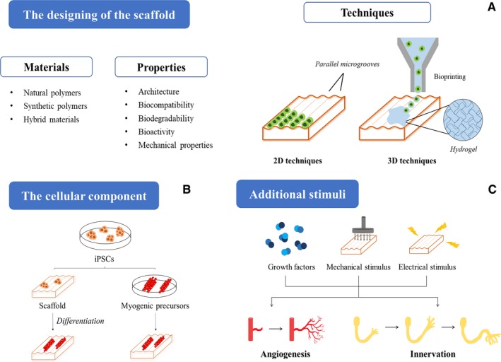
Both volumetric muscle loss (VML) and muscle degenerative diseases lead to an important decrease in skeletal muscle mass, condition that nowadays lacks an optimal treatment. This issue has driven towards an increasing interest in new strategies in tissue engineering, an emerging field that can offer very promising approaches. In addition, the discovery of induced pluripotent stem cells (iPSCs) has completely revolutionized the actual view of personalized medicine, and their utilization in skeletal muscle tissue engineering could, undoubtedly, add myriad benefits. In this review, we want to provide a general vision of the basic aspects to consider when engineering skeletal muscle tissue using iPSCs. Specifically, we will focus on the three main pillars of tissue engineering: the scaffold designing, the selection of the ideal cell source and the addition of factors that can enhance the resemblance with the native tissue.

Keywords: biomaterials; iPS-skeletal muscle; iPSCs; induced pluripotent stem cells; personalized medicine; regenerative medicine; scaffold; tissue engineering; volumetric muscle loss.


Conflict of interest statement

The authors confirm that there is no conflict of interest.

Figures

Figure 1
Figure 1
Skeletal muscle tissue. A, Skeletal muscle tissue comprises several bundles of aligned myofibres, each one containing thousands of myofibrils. In the image it is detailed the ultrastructure of the sarcomere, which is the final responsible of the contraction of the muscle due to the movement of myosin and actin myofilaments. B, The repair process of skeletal muscle is divided into three overlapping phases: destruction/inflammation, repair and remodelling. In the first stage, inflammatory cells are recruited to the damaged zone, secreting cytokines and growth factors which attract satellite cells. Then, adjacent blood vessels and nerves invade the healing area, and fibro/adipogenic progenitors co‐operate to form the provisional scar tissue. Finally, new myofibres are formed, and the repairing process concludes when contractile force is recovered
Figure 2
Figure 2
Main applications of patient‐derived iPSCs. Somatic cells from a patient can be reprogrammed using the Yamanaka factors (c‐Myc, Klf4, Sox2 and Oct3/4) into iPSCs. These generated iPSCs can be differentiated into the specific cell type affected in the disease, a step absolutely essential to create a cellular model. The potential applications of those models include drug screening, the elucidation of the pathophysiological mechanisms of the disease and even the discovery of new therapeutic approaches. Finally, all of this can lead to the development of a clinical trial, which could have a very positive impact on the patients themselves
Figure 3
Figure 3
Schematic representation of the three main pillars of skeletal muscle tissue engineering based on iPSCs. A, The scaffolds should present the right properties to ensure the correct engineering of the tissue, and they can be fabricated using natural, synthetic or hybrid polymers. In the image it is highlighted the main two‐dimensional and three‐dimensional techniques that can be employed for the generation of the scaffolds. B, iPSCs can be implemented to this technology following two strategies: either being seeded directly into the scaffold and then differentiated there, or cultivated in the scaffold once differentiated into myogenic precursors. C Finally, with the purpose of achieving vascularization and innervation in the construct, it is possible the addition of bioactive factors, as well as the implementation of mechanical or electrical stimuli

References

    1. Ostrovidov S, Hosseini V, Ahadian S, et al. Skeletal muscle tissue engineering: methods to form skeletal myotubes and their applications. Tissue Eng Part B Rev. 2014;20:403‐436. - PMC - PubMed
    1. Tabebordbar M, Wang ET, Wagers AJ. Skeletal muscle degenerative diseases and strategies for therapeutic muscle repair. Annu Rev Pathol Mech Dis. 2013;8:441‐475. - PubMed
    1. Huard J, Li Y, Fu FH. Muscle injuries and repair: current trends in research. J Bone Joint Surg Am. 2002;84:822‐832. - PubMed
    1. Yin H, Price F, Rudnicki MA. Satellite cells and the muscle stem cell niche. Physiol Rev. 2013;93:23‐67. - PMC - PubMed
    1. Turner NJ, Badylak SF. Regeneration of skeletal muscle. Cell Tissue Res. 2012;347:759‐774. - PubMed

Publication types

MeSH terms