

Review
doi: 10.1259/bjr.20180670.
Epub 2019 May 14.
Realizing the potential of magnetic resonance image guided radiotherapy in gynaecological and rectal cancer
Affiliations
- PMID: 30933550
- PMCID: PMC6592079
- DOI: 10.1259/bjr.20180670
Item in Clipboard
Review
Realizing the potential of magnetic resonance image guided radiotherapy in gynaecological and rectal cancer
Br J Radiol.
2019 Jun.
Abstract
CT-based radiotherapy workflow is limited by poor soft tissue definition in the pelvis and reliance on rigid registration methods. Current image-guided radiotherapy and adaptive radiotherapy models therefore have limited ability to improve clinical outcomes. The advent of MRI-guided radiotherapy solutions provides the opportunity to overcome these limitations with the potential to deliver online real-time MRI-based plan adaptation on a daily basis, a true "plan of the day." This review describes the application of MRI guided radiotherapy in two pelvic tumour sites likely to benefit from this approach.
Figures
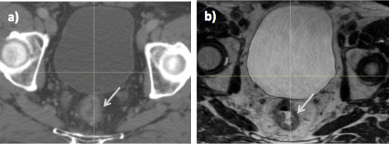
Radiotherapy planning imaging in a male patient with T3N1 rectal cancer; (a) CT and (b) MRI. On MRI, the tumour (arrow) is easily differentiated from normal rectum, which is not possible on CT.
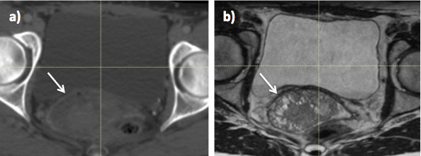
Radiotherapy planning imaging in Stage 2B cervix cancer (a) CT and (b) MRI. On MRI, the cervix tumour (arrow) is easily differentiated from normal bladder and rectum, which is not possible on CT.
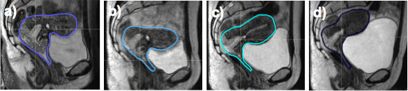
Changes in clinical target volume position during cervix radiotherapy as seen on MRI at (a) week 0, (b) week 2, (c) week 3 and (d) week 4.
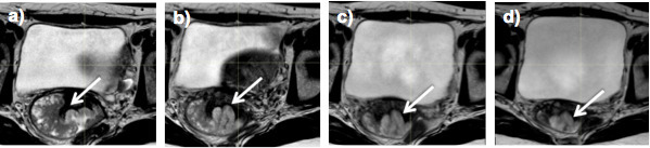
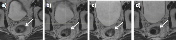
Changes in cervix tumour volume (arrow), as seen on weekly MRI during treatment at (a) week 0, (b) week 2, (c) week 3 and (d) week 4.
Changes in rectal tumour volume (arrow), as seen on weekly MRI during treatment at (a) week 0, (b) week 2, (c) week 3 and (d) week 4.
References
-
- Laursen LV , Elstrøm UV , Vestergaard A , Muren LP , Petersen JB , Lindegaard JC , et al. . Residual rotational set-up errors after daily cone-beam CT image guided radiotherapy of locally advanced cervical cancer . Radiother Oncol 2012. ; 105 : 220 – 5 . doi: 10.1016/j.radonc.2012.08.012 - DOI - PubMed
-
- Roeske JC , Lujan A , Rotmensch J , Waggoner SE , Yamada D , Mundt AJ . Intensity-modulated whole pelvic radiation therapy in patients with gynecologic Malignancies . International Journal of Radiation Oncology*Biology*Physics 2000. ; 48 : 1613 – 21 . doi: 10.1016/S0360-3016(00)00771-9 - DOI - PubMed
-
- Duthoy W , De Gersem W , Vergote K , Boterberg T , Derie C , Smeets P , et al. . Clinical implementation of intensity-modulated Arc therapy (IMAT) for rectal cancer . International Journal of Radiation Oncology*Biology*Physics 2004. ; 60 : 794 – 806 . doi: 10.1016/j.ijrobp.2004.04.016 - DOI - PubMed
-
- van Herk M , Remeijer P , Rasch C , Lebesque JV . The probability of correct target dosage: dose-population histograms for deriving treatment margins in radiotherapy . International Journal of Radiation Oncology*Biology*Physics 2000. ; 47 : 1121 – 35 . doi: 10.1016/S0360-3016(00)00518-6 - DOI - PubMed
