. 2019 Jul;33(7):8110-8124.
doi: 10.1096/fj.201802488R. Epub 2019 Apr 1.

Natural disease history of the D2 -mdx mouse model for Duchenne muscular dystrophy

Affiliations

Natural disease history of the D2 -mdx mouse model for Duchenne muscular dystrophy

Maaike van Putten et al. FASEB J. 2019 Jul.

Abstract

The C57BL/10ScSn-Dmdmdx/J (BL10-mdx) mouse has been the most commonly used model for Duchenne muscular dystrophy (DMD) for decades. Their muscle dysfunction and pathology is, however, less severe than in patients with DMD, which complicates preclinical studies. Recent discoveries indicate that disease severity is exacerbated when muscular dystrophy mouse models are generated on a DBA2/J genetic background. Knowledge on the natural history of animal models is pivotal for high-quality preclinical testing. However, for BL10-mdx mice on a DBA2/J background (D2-mdx), limited data are available. We addressed this gap in the natural history knowledge. First, we compared histopathological aspects in skeletal muscles of young D2-mdx, BL10-mdx, and wild-type mice. Pathology was more pronounced in D2-mdx mice and differed in severity between muscles within individuals. Secondly, we subjected D2-mdx mice to a functional test regime for 34 weeks and identified that female D2-mdx mice outperform severely impaired males, making females less useful for functional preclinical studies. Direct comparisons between 10- and 34-wk-old D2-mdx mice revealed that disease pathology ameliorates with age. Heart pathology was progressive, with some features already evident at a young age. This natural history study of the D2-mdx mouse will be instrumental for experimental design of future preclinical studies.-Van Putten, M., Putker, K., Overzier, M., Adamzek, W. A., Pasteuning-Vuhman, S., Plomp, J. J., Aartsma-Rus, A. Natural disease history of the D2-mdx mouse model for Duchenne muscular dystrophy.

Keywords: calcification; fibrosis; muscle function; pathology; regeneration.


Conflict of interest statement

The authors are grateful for the experimental help of Davy van de Vijver, Mandy Kater, and Joris van der Hurk [Leiden University Medical Centre (LUMC)]. This work was supported by funding from Duchenne Parent Project, French Muscular Dystrophy Association (AFM) Telethon (Grant 20251) and ZonMw (Grant 113302001) for researchers’ salaries. A.A.-R. is employed by Leiden University Medical Center (LUMC), which has patents on exon skipping technology, some of which has been licensed to BioMarin and subsequently sublicensed to Sarepta. As co-inventor of some of these patents, A.A.-R. is entitled to a share of royalties. A.A.-R. is also an ad hoc consultant for PTC Therapeutics, Summit PLC, Alpha Anomeric, BioMarin Pharmaceuticals, Eisai, Global Guidepoint and GLG consultancy, Grunenthal, Wave, and BioClinica. A.A.-R. was also a member of the Duchenne Network Steering Committee (BioMarin) and a member of the scientific advisory boards of ProQR, MirrX Therapeutics, and Philae Pharmaceuticals. Remuneration for these activities is paid to LUMC. LUMC also received speaker honoraria from PTC Therapeutics and BioMarin Pharmaceuticals and funding for contract research from Italpharmaco and Alpha Anomeric. The remaining authors declare no conflicts of interest.

Figures

Figure 1
Figure 1
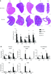
Histopathology of 10-wk-old mice. A) Representative H&E staining of 5 muscles belonging to 1 individual D2-mdx and BL10-mdx male. Scale bars, 1000 μm. Pathology consisted of necrosis, regenerated fibers, fibrosis, inflammation, and calcification and was more severe in D2-mdx mice. Differences between muscles were observed with the tibialis anterior being the least and diaphragm the most severely affected muscle in both dystrophic strains. B) Expression analysis of genes involved in fibrosis (Col1a1, Ctgf), inflammation (Cd68, Lgals3), and fat infiltration (Pparγ). Dia, diaphragm; gas, gastrocnemius; qua, quadriceps; ta, tibialis anterior; tri, triceps. Values represent means ± sd; n = 6 males/strain. #Significant difference compared with all other genotypes; *P < 0.05.
Figure 2
Figure 2
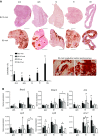
Myofiber calcification is extensive in 10-wk-old D2-mdx males. A) Representative Alizarin Red staining of 5 muscles belonging to the same D2-mdx and BL10-mdx males as shown in Fig. 1A. Scale bars, 1000 μm. Calcifications stain dark red and were primarily observed in D2-mdx mice. Longitudinal sections reveal that the calcified areas involve the entire myofiber and are contained within the fiber membrane. Quantifications revealed differences in severity between muscles. B) Expression analysis of genes involved in osteogenesis. Dia, diaphragm; gas, gastrocnemius; qua, quadriceps; ta, tibialis anterior; tri, triceps; n = 6 males/strain. Values represent means ± sd. #Significant difference compared with all other genotypes; *P < 0.05.
Figure 5
Figure 5
Histopathology is ameliorated in 34-wk-old D2-mdx mice. A) Gastrocnemius/body weight (GC/BW) ratio was smaller in D2-mdx mice. B) Representative H&E and Alizarin Red staining of the gastrocnemius and diaphragm belonging to a 10- and 34-wk-old D2-mdx male. The transient nature of the calcifications is clearly visible. Scale bars, 1000 μm. C, D) Overall pathology (C) and calcifications (D) were less pronounced in the older mice; n = 6 males/strain, except for 34-wk-old D2-mdx mice, which consisted of n = 10 males. Values represent means ± sd. #Statistically significant difference compared with all other genotypes; ***P < 0.0001.
Figure 3
Figure 3
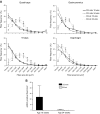
Regenerative capacity is not severely affected in 10-wk-old D2-mdx males. A) Fiber-size distributions were assessed for quadriceps, gastrocnemius, triceps, and diaphragm of the 4 strains. Overall, D2-mdx and BL10-mdx mice had significantly smaller fibers compared with WT strains. D2-mdx mice had a larger proportion of small fibers in gastrocnemius and triceps than BL10-mdx mice. X-axis labeling of quadriceps, triceps, and gastrocnemius are identical. B) Representative H&E, Alizarin Red, and eMHC stainings of consecutive sections of a D2-mdx and a BL10-mdx male quadriceps. Scale bars, 100 μm. Symbols indicate corresponding fibers. eMHC-positive fibers colocalized to areas of inflammation and calcification. C) eMHC-positive fibers were manually counted on an entire cross section. Abundance did not differ between D2-mdx and BL10-mdx males. D) Expression analysis of genes involved in regeneration. Dia, diaphragm; gas, gastrocnemius; tri, triceps; n = 6 males/strain. Values represent means ± sem (A) and mean ± sd (BD). #Significant difference compared with all other genotypes; *P < 0.05.
Figure 4
Figure 4
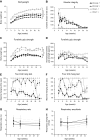
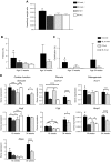
A gender and age comparison for D2-mdx and D2-WT mice shows that muscle function is severely affected in D2-mdx males, but not females, and deteriorates with age. A) D2-mdx mice were lighter than wild types. B) CK levels were elevated in young D2-mdx mice and dropped with age. C, D) Forelimb grip strength (C) was impaired in D2-mdx mice, but when normalized to body weight (D), this difference lost its significance. E, F) Performance in the 2 limb hanging test (E) and 4 limb hanging test (F) was severely impaired in D2-mdx males but not in females. G) Respiratory rate was decreased in D2-mdx mice, although this did not reach significance. H) Respiratory amplitude normalized to body weight showed a nonsignificant increase in D2-mdx mice. For G and H, significance was reached when data of both genders were pooled; n = 10 D2-mdx mice and n = 6 D2-WT mice of both genders. Values represent means ± sem. $Significant difference compared with all other groups; significant drop with age in D2-mdx mice; *P < 0.05.
Figure 6
Figure 6
Fiber-size and regeneration in 34-wk-old D2-mdx mice. A) Fiber-size distributions were similar between younger and older D2-mdx mice for quadriceps and triceps. X-axis labeling of quadriceps, triceps, and gastrocnemius are identical. B) eMHC-positive fibers were only occasionally observed in old D2-mdx mice. Values represent means ± sd.
Figure 7
Figure 7
Pathology of the heart of D2-mdx and BL10-mdx mice at 10 and 34 wk of age. A) D2-mdx males, but not females, suffered from cardiac hypertrophy at the age of 34 wk. B) Despite large individual variation, D2-mdx mice had marginally more fibrotic lesions from 10 wk of age onwards compared with BL10-mdx mice. C) In contrast to BL10-mdx mice, D2-mdx mice had calcifications in the heart at 10 wk of age. D) Expression analysis of genes involved in cardiac function (Serca2a, Vegf, and Nppa), fibrosis (Col1a1, Ctgf) and osteogenesis (Acvr1, Bmp2); n = 6 males/strain, except for 34-wk-old D2-mdx mice, which consisted of n = 10 males. Values represent means ± sd. #Statistically significant difference compared with all other genotypes; *P < 0.05, **P < 0.01, ***P < 0.001, ****P < 0.0001.

References

    1. Mendell J. R., Rodino-Klapac L. R., Sahenk Z., Roush K., Bird L., Lowes L. P., Alfano L., Gomez A. M., Lewis S., Kota J., Malik V., Shontz K., Walker C. M., Flanigan K. M., Corridore M., Kean J. R., Allen H. D., Shilling C., Melia K. R., Sazani P., Saoud J. B., Kaye E. M.; Eteplirsen Study Group (2013) Eteplirsen for the treatment of Duchenne muscular dystrophy. Ann. Neurol. 74, 637–647 - PubMed
    1. Moat S. J., Bradley D. M., Salmon R., Clarke A., Hartley L. (2013) Newborn bloodspot screening for Duchenne muscular dystrophy: 21 years experience in Wales (UK). Eur. J. Hum. Genet. 21, 1049–1053 - PMC - PubMed
    1. Straub V., Balabanov P., Bushby K., Ensini M., Goemans N., De Luca A., Pereda A., Hemmings R., Campion G., Kaye E., Arechavala-Gomeza V., Goyenvalle A., Niks E., Veldhuizen O., Furlong P., Stoyanova-Beninska V., Wood M. J., Johnson A., Mercuri E., Muntoni F., Sepodes B., Haas M., Vroom E., Aartsma-Rus A. (2016) Stakeholder cooperation to overcome challenges in orphan medicine development: the example of Duchenne muscular dystrophy. Lancet Neurol. 15, 882–890 - PubMed
    1. Emery A. E. (2002) The muscular dystrophies. Lancet 359, 687–695 - PubMed
    1. Kim S., Campbell K. A., Fox D. J., Matthews D. J., Valdez R.; MD STARnet (2015) Corticosteroid treatments in males with Duchenne muscular dystrophy: treatment duration and time to loss of ambulation. J. Child Neurol. 30, 1275–1280 - PMC - PubMed

Publication types

MeSH terms

LinkOut - more resources