. 2019 Mar 25;11(3):700.
doi: 10.3390/nu11030700.

Protective Effects of CISD2 and Influence of Curcumin on CISD2 Expression in Aged Animals and Inflammatory Cell Model

Affiliations

Protective Effects of CISD2 and Influence of Curcumin on CISD2 Expression in Aged Animals and Inflammatory Cell Model

Chai-Ching Lin et al. Nutrients. .

Abstract

Background: Inflammation and mitochondrial dysfunction have been linked to trauma, neurodegeneration, and aging. Impairment of CISD2 expression may trigger the aforementioned pathological conditions in neural cells. We previously reported that curcumin attenuates the downregulation of CISD2 in animal models of spinal cord injury and lipopolysaccharide (LPS)-treated neuronal cells. In this study, we investigate (1) the role of CISD2 and (2) how curcumin regulates CISD2 in the aging process.

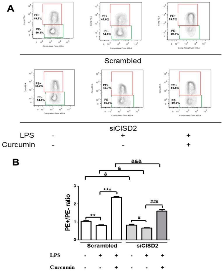
Materials and methods: The serial expression of CISD2 and the efficacy of curcumin treatment were evaluated in old (104 weeks) mice and long-term cultures of neural cells (35 days in vitro, DIV). LPS-challenged neural cells (with or without siCISD2 transfection) were used to verify the role of curcumin on CISD2 underlying mitochondrial dysfunction.

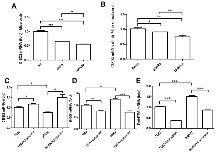
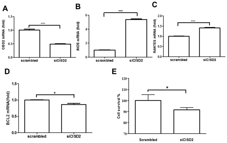
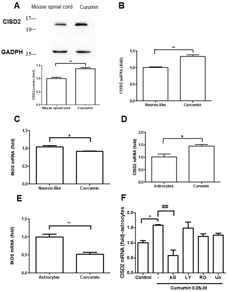
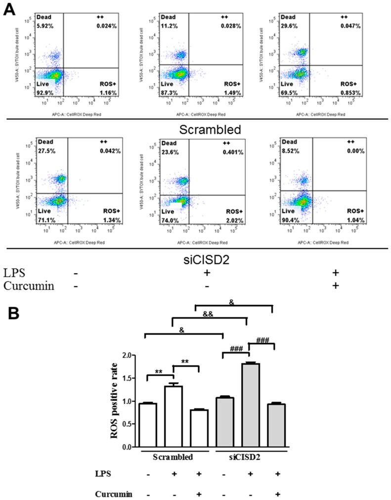
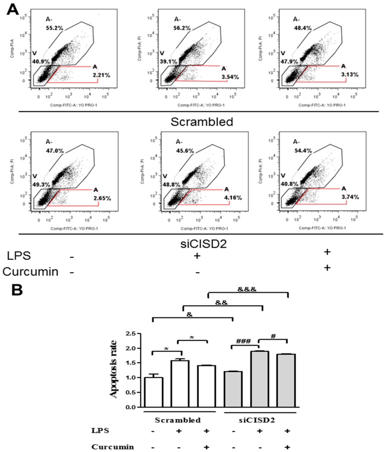
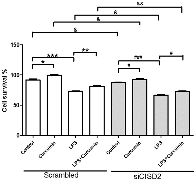
Results: In the brain and spinal cord of mice aged P2, 8, 25, and 104 weeks, we observed a significant decrease in CISD2 expression with age. Curcumin treatment in vivo and in vitro was shown to upregulate CISD2 expression; attenuate inflammatory response in neural cells. Moreover, curcumin treatment elevated CISD2 expression levels and prevented mitochondrial dysfunction in LPS-challenged neural cells. The beneficial effects of curcumin in either non-stressed or LPS-challenged cells that underwent siCISD2 transfection were significantly lower than in respective groups of cells that underwent scrambled siRNA-transfection.

Conclusions: We hypothesize that the protective effects of curcumin treatment in reducing cellular inflammation associated trauma, degenerative, and aging processes can be partially attributed to elevated CISD2 expression. We observed a reduction in the protective effects of curcumin against injury-induced inflammation and mitochondrial dysfunction in cells where CISD2 expression was reduced by siCISD2.

Keywords: CISD2; CISD2-dependent manner; aging; curcumin; neurodegeneration; trauma.


Conflict of interest statement

No competing financial interests exist. The authors declare no conflict of interest.

Figures

Figure 1
Figure 1
Curcumin upregulated CISD2 expression in aged mice in vivo. (A,B) In vivo mouse model of aging. Results of CISD2 mRNA expression in the brains (A) and spinal cords (B) of mice at P2, 8, 25 and 104 weeks (n = 4 in each group). Curcumin enhanced CISD2 expression in a long-term culture of neural cells in vitro. Results of mRNA expression of CISD2 (C), iNOS (D), and RANTES (E) in the neural cells of mice with or without 1 μM curcumin treatment at 7 DIV and 35 DIV. Vertical bars indicate the mean ± (standard error of the mean (SEM)) of mRNA expression (n = 3). * p < 0.05, ** p < 0.01, and *** p < 0.001 indicate differences of statistical significance.
Figure 2
Figure 2
Knockdown of CISD2 expression in neural cells produced inflammatory responses, promoted apoptosis, and decreased cell viability. Results of mRNA expression of CISD2 (A), iNOS (B), RANTES (C), and BCL2 (D) in neural cells with or without siCISD2 transfection. (E) Cell viability was measured using alamarBlue® assay. Vertical bars indicate the mean ± (SEM) of mRNA expression (n = 3). * p < 0.05, and *** p < 0.001 indicate differences of statistical significance.
Figure 3
Figure 3
Curcumin upregulated CISD2 protein expression in vivo. (A) CISD2 protein expression in mouse spinal cords following intraperitoneal administration of curcumin at a concentration of 40 mg/kg. The upper panel presents the results of immunoblotting analysis of CISD2 (15 kDa) with GAPDH (37 kDa) serving as an internal control. The lower panel presents the mean ± (SEM) of CISD2/GAPDH band intensity as a ratio of the results from the control group (n = 4). Curcumin enhanced CISD2 mRNA expression in vitro via JAK/STAT signaling pathways. mRNA expression of CISD2 (B) and iNOS (C) in neuron-like cells (SH-SY5Y). mRNA expression levels of CISD2 (D) and iNOS (E) in astrocyte culture. (F) CISD2 mRNA expression following treatment with curcumin and the inhibition of JAK/STAT signaling pathways in non-stimulated astrocytes: AG490: JAK/STAT inhibitor, LY294002: PI3K inhibitor, RO318220: PKC inhibitor, U0126: MAPK inhibitor. Vertical bars indicate the mean + (SEM) of mRNA expression (n = 3). * p < 0.05, and ##,** p < 0.01 indicate differences of statistical significance.
Figure 4
Figure 4
Mitochondrial membrane potential [DeltaPsi(m)] of lipopolysaccharide (LPS)-challenged neural cells (with or without curcumin treatment), as determined using JC-1 staining and flow cytometry with or without CISD2 knockdown. Cells presenting a decrease in DeltaPsi(m) and impending cellular apoptosis are indicated by monomer green JC-1 fluorescence, compared to normal cells with intact DeltaPsi(m) (indicated by j-aggregated red fluorescence). All groups of cells were evaluated in terms of the ratio of PE+ to PE−. (A) Representative results of flow cytometry for the following groups of cells: (i) scramble RNA-transfected control cells untreated with LPS; (ii) scramble RNA-transfected, LPS-challenged cells, untreated with curcumin; (iii) scramble RNA-transfected, LPS-challenged cells following curcumin treatment; (iv) siCID2-transfected control cells without LPS or curcumin treatment; (v) siCID2-transfected, LPS-challenged cells without curcumin treatment; (vi) siCID2-transfected, LPS-challenged cells, treated with curcumin. (B) Bars indicate the mean ± SEM (n = 3). #,& p < 0.05, ** p < 0.01, and ###,&&&,*** p < 0.001 indicate differences of statistical significance.
Figure 5
Figure 5
Reactive oxygen species (ROS) formation in LPS-challenged neural cells (with or without curcumin treatment), as determined by CellROX® Deep Red/SYTOX® Blue Dead Cell stain and flow cytometry with or without CISD2 knockdown. RegionLive: vital cells; Region Dead: dead cells; Region++: dead cells with accumulated ROS; Region ROS+: vital cells with ROS formation. SYTOX ® Blue Dead Cell stain was used to distinguish vital cells from dead cells. ROS+ cells of all groups were analyzed using flow cytometry (A). Representative flow cytometry results from all groups. (B) Bars indicate the mean ± SEM (n = 3). & p < 0.05, &&,** p < 0.01, and ### p < 0.001 indicate differences of statistical significance.
Figure 6
Figure 6
Apoptosis of LPS-challenged neural cells with or without curcumin treatment, as determined using YO-PRO-1/PI double staining and flow cytometry with or without CISD2 knockdown. YO-PRO-1 serves as a marker for apoptosis. PI refers to propidium iodide, which is used as a marker for necrotic cells. Region A-: non-living cells without early apoptosis; Region V: vital cells; Region A: early apoptotic cells. Early apoptotic cells with positive YO-PRO-1 and negative PI staining were calculated and compared among all groups (A). Representative flow cytometry results from all groups (B). Bars indicate the mean ± SEM (n = 3). &,#,* p < 0.05, && p < 0.01, and &&&,### p < 0.001 indicate differences of statistical significance.
Figure 7
Figure 7
Curcumin enhanced cell viability and attenuated LPS-challenge-driven cell death in non-stressed and LPS-challenged neural cells. The knockdown of CISD2 expression was shown to reduce the protective effects of curcumin. The eight groups of cells included the following: (i) scrambled RNA-transfected control cells, untreated with LPS and curcumin; (ii) scrambled RNA-transfected cells treated with curcumin; (iii) scramble RNA-transfected, LPS-challenged cells, without curcumin treatment; (iv) scramble RNA-transfected, LPS-challenged cells treated with curcumin; (v) siCID2-transfected control cells without LPS or curcumin treatment; (vi) siCID2-transfected cells treated with curcumin; (vii) siCID2-transfected, LPS-challenged cells without curcumin treatment; (viii) siCID2-transfected, LPS-challenged cells, treated with curcumin. Cell viability was measured using alamarBlue® assay. Vertical bars indicate the mean ± SEM (n = 3). #,&,* p < 0.05, &&,** p < 0.01, and ###,*** p < 0.001 indicate differences of statistical significance.

References

    1. Kline K.A., Bowdish D.M. Infection in an aging population. Curr. Opin. Microbiol. 2016;29:63–67. doi: 10.1016/j.mib.2015.11.003. - DOI - PubMed
    1. Maragakis N.J., Rothstein J.D. Mechanisms of Disease: Astrocytes in neurodegenerative disease. Nat. Clin. Pract. Neurol. 2006;2:679–689. doi: 10.1038/ncpneuro0355. - DOI - PubMed
    1. Hickman S., Izzy S., Sen P., Morsett L., El K.J. Microglia in neurodegeneration. Nat. Neurosci. 2018;21:1359–1369. doi: 10.1038/s41593-018-0242-x. - DOI - PMC - PubMed
    1. Ravari A., Mirzaei T., Kennedy D., Kazemi A.M. Chronoinflammaging in Alzheimer; A systematic review on the roles of toll like receptor 2. Life Sci. 2017;171:16–20. doi: 10.1016/j.lfs.2017.01.003. - DOI - PubMed
    1. Navarro A., Boveris A. Brain mitochondrial dysfunction in aging, neurodegeneration, and Parkinson’s disease. Front. Aging Neurosci. 2010;2:34. doi: 10.3389/fnagi.2010.00034. - DOI - PMC - PubMed

MeSH terms