. 2019 Mar 25;20(6):1490.
doi: 10.3390/ijms20061490.

Horse Oil Mitigates Oxidative Damage to Human HaCaT Keratinocytes Caused by Ultraviolet B Irradiation

Affiliations

Horse Oil Mitigates Oxidative Damage to Human HaCaT Keratinocytes Caused by Ultraviolet B Irradiation

Mei Jing Piao et al. Int J Mol Sci. .

Abstract

Horse oil products have been used in skin care for a long time in traditional medicine, but the biological effects of horse oil on the skin remain unclear. This study was conducted to evaluate the protective effect of horse oil on ultraviolet B (UVB)-induced oxidative stress in human HaCaT keratinocytes. Horse oil significantly reduced UVB-induced intracellular reactive oxygen species and intracellular oxidative damage to lipids, proteins, and DNA. Horse oil absorbed light in the UVB range of the electromagnetic spectrum and suppressed the generation of cyclobutane pyrimidine dimers, a photoproduct of UVB irradiation. Western blotting showed that horse oil increased the UVB-induced Bcl-2/Bax ratio, inhibited mitochondria-mediated apoptosis and matrix metalloproteinase expression, and altered mitogen-activated protein kinase signaling-related proteins. These effects were conferred by increased phosphorylation of extracellular signal-regulated kinase 1/2 and decreased phosphorylation of p38 and c-Jun N-terminal kinase 1/2. Additionally, horse oil reduced UVB-induced binding of activator protein 1 to the matrix metalloproteinase-1 promoter site. These results indicate that horse oil protects human HaCaT keratinocytes from UVB-induced oxidative stress by absorbing UVB radiation and removing reactive oxygen species, thereby protecting cells from structural damage and preventing cell death and aging. In conclusion, horse oil is a potential skin protectant against skin damage involving oxidative stress.

Keywords: apoptosis; horse oil; oxidative stress; ultraviolet B radiation.


Conflict of interest statement

The authors declare no conflict of interest.

Figures
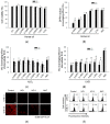
Figure 1
Figure 1
Horse oil can scavenge stress-induced ROS. (a) Horse oil (HO-I or HO-II) was added at the indicated final concentrations. After 24 h, cell viability was measured by MTT assay. (b) DPPH radical levels were measured spectrophotometrically at 520 nm. *, # p < 0.05 vs. non-treated control cells. Intracellular ROS levels generated by (c) H2O2 or (d) UVB radiation were detected by spectrofluorometry after H2DCFDA staining. NAC served as a positive control. * p < 0.05 vs. H2O2 alone, and # p < 0.05 vs. UVB alone. Cells were treated with 0.312% horse oil for 1 h, and then with UVB radiation at 30 mJ/cm2. Next, the cells were incubated for 24 h, and intracellular ROS were detected by (e) confocal microscopy (Scale bar = 10 μm) and (f) flow cytometry after H2DCFDA staining.
Figure 2
Figure 2
Horse oil mitigates UVB-induced oxidative damage to cellular macromolecules. Cells were treated with horse oil or NAC for 1 h and then exposed to UVB radiation. After incubation, lipid peroxidation was assayed by measuring (a) the levels of 8-isoprostane secreted into the culture medium or (b) DPPP-stained cells were detected by confocal microscopy and quantified (Scale bar=10 μm). (c) Protein oxidation was assayed by measuring protein carbonylation. (d) DNA damage was assessed by the comet assay. Representative images and the percentage of total cellular DNA in comet tails are shown (Scale bar=10 μm). * p < 0.05 vs. control, and # p < 0.05 vs. UVB-irradiated cells. (e) 8-OxoG detected by the binding of avidin-TRITC was visualized by confocal microscopy (Scale bar=10 μm). DNA was extracted and analyzed by (f) ELISA and (g) immunocytochemistry using an antibody against CPDs. DAPI was used to stain the nuclei (Scale bar = 10 μm). * p < 0.05 vs. control, and # p < 0.05 vs. UVB-irradiated cells.
Figure 2
Figure 2
Horse oil mitigates UVB-induced oxidative damage to cellular macromolecules. Cells were treated with horse oil or NAC for 1 h and then exposed to UVB radiation. After incubation, lipid peroxidation was assayed by measuring (a) the levels of 8-isoprostane secreted into the culture medium or (b) DPPP-stained cells were detected by confocal microscopy and quantified (Scale bar=10 μm). (c) Protein oxidation was assayed by measuring protein carbonylation. (d) DNA damage was assessed by the comet assay. Representative images and the percentage of total cellular DNA in comet tails are shown (Scale bar=10 μm). * p < 0.05 vs. control, and # p < 0.05 vs. UVB-irradiated cells. (e) 8-OxoG detected by the binding of avidin-TRITC was visualized by confocal microscopy (Scale bar=10 μm). DNA was extracted and analyzed by (f) ELISA and (g) immunocytochemistry using an antibody against CPDs. DAPI was used to stain the nuclei (Scale bar = 10 μm). * p < 0.05 vs. control, and # p < 0.05 vs. UVB-irradiated cells.
Figure 3
Figure 3
Horse oil attenuates UVB-induced apoptosis. HaCaT keratinocytes were treated with horse oil or NAC and exposed to UVB radiation 1 h later. Cells were then incubated for 24 h. Δψm was analyzed by (a) confocal microscopy after staining the cells with JC-1 (Scale bar = 10 μm). (b) DNA fragmentation was quantified by ELISA. (c) Apoptotic bodies (arrows) were observed in cells stained with Hoechst 33342 dye and quantified by fluorescence microscopy (Scale bar = 10 μm). (d) Cell viability following UVB radiation was determined by MTT assay. (e,f) Western blotting with antibodies specific for (e) PARP, caspase-9, caspase-3, Bax, Bcl-2, actin, (f) phospho-ERK1/2, phospho-p38, and phospho-JNK1/2, and actin, and the results were quantified (n = 3). * p < 0.05 vs. control, and # p < 0.05 vs. UVB-irradiated cells.
Figure 3
Figure 3
Horse oil attenuates UVB-induced apoptosis. HaCaT keratinocytes were treated with horse oil or NAC and exposed to UVB radiation 1 h later. Cells were then incubated for 24 h. Δψm was analyzed by (a) confocal microscopy after staining the cells with JC-1 (Scale bar = 10 μm). (b) DNA fragmentation was quantified by ELISA. (c) Apoptotic bodies (arrows) were observed in cells stained with Hoechst 33342 dye and quantified by fluorescence microscopy (Scale bar = 10 μm). (d) Cell viability following UVB radiation was determined by MTT assay. (e,f) Western blotting with antibodies specific for (e) PARP, caspase-9, caspase-3, Bax, Bcl-2, actin, (f) phospho-ERK1/2, phospho-p38, and phospho-JNK1/2, and actin, and the results were quantified (n = 3). * p < 0.05 vs. control, and # p < 0.05 vs. UVB-irradiated cells.
Figure 4
Figure 4
Horse oil absorbs UVB rays. The UVB absorption spectrum of horse oil was determined by UV scanning at 200–400 nm. The maximum absorbance of horse oil appeared at 271 nm, and the absorbance values of HO-I or HO-II were 0.2756 and 0.5067, respectively.
Figure 5
Figure 5
Horse oil reduces UVB-induced MMP expression and activation. (a) Western blot analysis of MMP-1, MMP-2, and MMP-9 protein expression and quantitative data are shown (n = 3). (b) Active MMP-1 was quantified in the culture supernatants. Cells were incubated in serum-free medium to eliminate interference from MMP-1 in the serum. * p < 0.05 vs. control, and # p < 0.05 vs. UVB-irradiated cells. (c) AP-1 binding to the MMP-1 promoter was assessed by ChIP assay.

References

    1. Brooker E.G., Shorland F.B. Studies on the composition of horse oil. 1. Composition of horse oil in relation to the depot fats of other pasture-fed animals. Biochem. J. 1950;46:80–85. doi: 10.1042/bj0460080. - DOI - PMC - PubMed
    1. Al-Shudiefat A.A., Sharma A.K., Bagchi A.K., Dhingra S., Singal P.K. Oleic acid mitigates TNF-α-induced oxidative stress in rat cardiomyocytes. Mol. Cell. Biochem. 2013;372:75–82. doi: 10.1007/s11010-012-1447-z. - DOI - PubMed
    1. Guzmán D.C., Brizuela N.O., Herrera M.O., Olguín H.J., García E.H., Peraza A.V., Mejía G.B. Oleic acid protects against oxidative stress exacerbated by cytarabine and doxorubicin in rat brain. Anticancer Agents Med. Chem. 2016;16:1491–1495. doi: 10.2174/1871520615666160504093652. - DOI - PubMed
    1. Basiricò L., Morera P., Dipasquale D., Tröscher A., Bernabucci U. Comparison between conjugated linoleic acid and essential fatty acids in preventing oxidative stress in bovine mammary epithelial cells. J. Dairy Sci. 2017;100:2299–2309. doi: 10.3168/jds.2016-11729. - DOI - PubMed
    1. Kim H., Youn K., Yun E.Y., Hwang J.S., Jeong W.S., Ho C.T., Jun M. Oleic acid ameliorates Aβ-induced inflammation by downregulation of COX-2 and iNOS via NFκB signaling pathway. J. Funct. Foods. 2015;14:1–11. doi: 10.1016/j.jff.2015.01.027. - DOI