Review
. 2019 Mar 30;11(4):456.
doi: 10.3390/cancers11040456.

TRAILblazing Strategies for Cancer Treatment

Affiliations
Review

TRAILblazing Strategies for Cancer Treatment

Anna-Laura Kretz et al. Cancers (Basel). .

Abstract

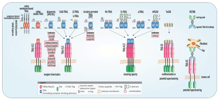
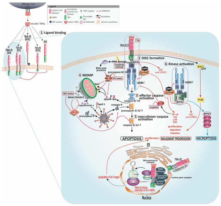
In the late 1990s, tumor necrosis factor (TNF)-related apoptosis-inducing ligand (TRAIL), a member of the TNF-family, started receiving much attention for its potential in cancer therapy, due to its capacity to induce apoptosis selectively in tumour cells in vivo. TRAIL binds to its membrane-bound death receptors TRAIL-R1 (DR4) and TRAIL-R2 (DR5) inducing the formation of a death-inducing signalling complex (DISC) thereby activating the apoptotic cascade. The ability of TRAIL to also induce apoptosis independently of p53 makes TRAIL a promising anticancer agent, especially in p53-mutated tumour entities. Thus, several so-called TRAIL receptor agonists (TRAs) were developed. Unfortunately, clinical testing of these TRAs did not reveal any significant anticancer activity, presumably due to inherent or acquired TRAIL resistance of most primary tumour cells. Since the potential power of TRAIL-based therapies still lies in TRAIL's explicit cancer cell-selectivity, a desirable approach going forward for TRAIL-based cancer therapy is the identification of substances that sensitise tumour cells for TRAIL-induced apoptosis while sparing normal cells. Numerous of such TRAIL-sensitising strategies have been identified within the last decades. However, many of these approaches have not been verified in animal models, and therefore potential toxicity of these approaches has not been taken into consideration. Here, we critically summarise and discuss the status quo of TRAIL signalling in cancer cells and strategies to force tumour cells into undergoing apoptosis triggered by TRAIL as a cancer therapeutic approach. Moreover, we provide an overview and outlook on innovative and promising future TRAIL-based therapeutic strategies.

Keywords: TRAIL in cancer; TRAIL sensitising; TRAIL signalling; TRAIL-induced apoptosis.


Conflict of interest statement

The authors declare no conflict of interest.

Figures

Figure 2
Figure 2
Highly potent TRAIL receptor agonists (TRAs) and combinatorial approaches. Schematic illustration of native tumour-necrosis-factor-related apoptosis-inducing ligand (TRAIL) and potent TRAs evaluated in clinical studies or with promising results in preclinical experiments and their combination with anticancer approaches: native membrane-bound TRAIL is a protein of 281 amino acids (aa) comprising a cytoplasmic domain (aa 1–17), a transmembrane domain (aa 18–38), and an extracellular domain (aa 39–281). The receptor-binding domain (aa 114–281) is located in the extracellular portion [1] which is cleaved from the cell surface to form bell-shaped homo-trimers. Recombinant human TRAIL/Apo2L (dulanermin) is based on the amino acids 114–281 of the extracellular portion of TRAIL [4]. The patients’ benefit in clinical trials was merely marginal even combined with chemotherapeutic regimens such as FOLFIRI (folinic acid, fluorouracil, irinotecan) [93,94]. Tags at the N-terminus of the extracellular domain of TRAIL such as FLAG and poly-histidine (His) aid purification of the ligand and leucine zipper (LZ) and isoleucine zipper (iz) stabilize the trimers [3,120]. Circularly permuted TRAIL (CPT) consists of the N-terminal amino acids 121–135 linked to the C-terminal amino acids 135-281 of TRAIL via a flexible linker [121]. To achieve the fragment crystallizable region (Fc)-TRAIL fusion protein, the Fc portion of human immunoglobulin G1 (IgG1) was fused to the N-terminus of human TRAIL (aa 95 to 281) [122]. Single-chain TRAIL (scTRAIL) is assembled by 3 extracellular domains of TRAIL that are covalently connected by 2 short peptide sequences [123]. APG350 was developed by the fusion of scTRAIL and the Fc-part of a human IgG1 leading to six receptor binding sites per drug molecule [124]. TAS266 comprises four humanised high-affinity heavy chain domain (VHH) antibody fragments which enable clustering of four TRAIL-R2s [117]. The substance caused severe hepatoxicity in clinical phase I studies. The tetravalent fibroblast-activation protein (FAP)-TRAIL-R2 antibody RG7386, targets cancer-associated fibroblasts in the tumour stroma and TRAIL-R2 on tumour cells simultaneously thereby inducing higher-order clustering and apoptosis induction in vitro and animal models [125].
Figure 1
Figure 1
TRAIL signalling and sensitisation. Human tumour-necrosis-factor-related apoptosis-inducing ligand (TRAIL) can bind to four membrane-bound TRAIL receptors (TRAIL-Rs) and one soluble receptor ①. TRAIL-R1 and TRAIL-R2 can trigger apoptotic signals while the other receptors might serve as decoys and regulate apoptosis negatively. Cysteine-rich domains of the receptors are crucial for ligand binding. Following TRAIL binding to TRAIL-R1 or TRAIL-R2, the death-inducing signalling complex (DISC) is assembled ②. In type I cells, the signal of DISC-released caspase-8 is sufficient for activation of further downstream caspases and apoptosis induction ③, whereas the DISC signal is amplified via mitochondria in type II cells ④. Cleaved and thereby truncated Bid (tBid) activates the mitochondria-associated B-cell CLL/lymphoma 2 (Bcl-2)-family members Bcl-2-associated X protein (Bax) and Bcl-2 antagonist or killer (Bak) resulting in mitochondrial outer membrane permeabilisation (MOMP) and finally cytochrome c and second mitochondrial activator of caspases/direct inhibitor of apoptosis-binding protein with low isoelectric point (pI) (SMAC/DIABLO) release. The apoptosome comprising apoptotic protease activating factor-1 (Apaf-1), cytochrome c, and caspase-9, presents the activation platform for caspase-9 which leads eventually to the activation of executioner caspases ⑤. p53 is activated in response to stress signals such as DNA damage and promotes Bcl-2 homology (BH)3-only proteins resulting in MOMP. TRAIL-receptor-interaction can provoke the formation of a second cytosolic complex ⑥, retaining Fas-associated protein with death domain (FADD) and caspase-8 and recruiting receptor-interacting serine/threonine-protein kinase 1 (RIPK1), TNF receptor-associated factor 2 (TRAF2), and nuclear factor kappa-light-chain-enhancer of activated B cells (NF-κB) essential modifier (NEMO) [38,39,40]. TRAF2 recruits cellular inhibitor of apoptosis protein 1/2 (cIAP1/2) which in turn trigger the ubiquitination of RIPK1 and therefore recruitment of linear ubiquitin chain assembly complex (LUBAC) [41]. LUBAC poly-ubiquitinylates RIPK1. RIPK1 is the stimulus for tyrosine-protein kinase Src and signal transducer and activator of transcription 3 (STAT3) promoting migration and invasion [42]. Complex I and complex II induce NF-κB, p38 mitogen-activated protein kinase (p38 MAPK), c-JUN N-terminal kinase (JNK) and extracellular signal-regulated kinase (ERK) pathways. LUBAC is present in both complexes, responsible for caspase-8 activation and recruitment of the inhibitor of κB (IκB) kinase (IKK) complex, and consequently activation of NF-κB [41]. In case of blocked caspase-8, the necrosome is formed by the interaction of RIPK1 and RIPK3. Independently of FADD and complex I and II, the membrane-proximal domain (MPD) of TRAIL-R2 can activate Ras-related C3 botulinum toxin substrate 1 (Rac1) to promote migration and invasion [43]. TRAIL-R2 can also occur in the nucleus where it interacts with ribonucleoprotein complexes involved in the maturation of microRNAs (miRNAs) of the let-7 family. These miRNAs interact with and constrain mRNAs of several regulators of mitogenic pathways such as Ras and c-Myc thereby encouraging proliferation of tumour cells [44]. Further abbreviations: glycosylphosphatidylinositol (GPI), osteoprotegerin (OPG), death domain (DD).

References

    1. Wiley S.R., Schooley K., Smolak P.J., Din W.S., Huang C.P., Nicholl J.K., Sutherland G.R., Smith T.D., Rauch C., Smith C.A., et al. Identification and characterization of a new member of the TNF family that induces apoptosis. Immunity. 1995;3:673–682. doi: 10.1016/1074-7613(95)90057-8. - DOI - PubMed
    1. Pitti R.M., Marsters S.A., Ruppert S., Donahue C.J., Moore A., Ashkenazi A. Induction of apoptosis by Apo-2 ligand, a new member of the tumor necrosis factor cytokine family. J. Biol. Chem. 1996;271:12687–12690. doi: 10.1074/jbc.271.22.12687. - DOI - PubMed
    1. Walczak H., Miller R.E., Ariail K., Gliniak B., Griffith T.S., Kubin M., Chin W., Jones J., Woodward A., Le T., et al. Tumoricidal activity of tumor necrosis factor-related apoptosis-inducing ligand in vivo. Nat. Med. 1999;5:157–163. doi: 10.1038/5517. - DOI - PubMed
    1. Ashkenazi A., Pai R.C., Fong S., Leung S., Lawrence D.A., Marsters S.A., Blackie C., Chang L., McMurtrey A.E., Hebert A., et al. Safety and antitumor activity of recombinant soluble Apo2 ligand. J. Clin. Investig. 1999;104:155–162. doi: 10.1172/JCI6926. - DOI - PMC - PubMed
    1. Pan G., Ni J., Wei Y.F., Yu G., Gentz R., Dixit V.M. An antagonist decoy receptor and a death domain-containing receptor for TRAIL. Science (N. Y.) 1997;277:815–818. doi: 10.1126/science.277.5327.815. - DOI - PubMed