. 2019 Mar 31;20(7):1604.
doi: 10.3390/ijms20071604.

Transcriptomics and Immunological Analyses Reveal a Pro-Angiogenic and Anti-Inflammatory Phenotype for Decidual Endothelial Cells

Affiliations

Transcriptomics and Immunological Analyses Reveal a Pro-Angiogenic and Anti-Inflammatory Phenotype for Decidual Endothelial Cells

Chiara Agostinis et al. Int J Mol Sci. .

Abstract

Background: In pregnancy, excessive inflammation and break down of immunologic tolerance can contribute to miscarriage. Endothelial cells (ECs) are able to orchestrate the inflammatory processes by secreting pro-inflammatory mediators and bactericidal factors by modulating leakiness and leukocyte trafficking, via the expression of adhesion molecules and chemokines. The aim of this study was to analyse the differences in the phenotype between microvascular ECs isolated from decidua (DECs) and ECs isolated from human skin (ADMECs).

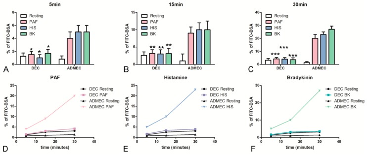
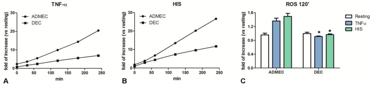
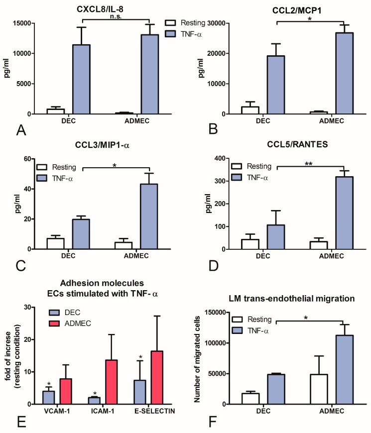
Methods: DECs and ADMECs were characterized for their basal expression of angiogenic factors and adhesion molecules. A range of immunological responses was evaluated, such as vessel leakage, reactive oxygen species (ROS) production in response to TNF-α stimulation, adhesion molecules expression and leukocyte migration in response to TNF-α and IFN-γ stimulation.

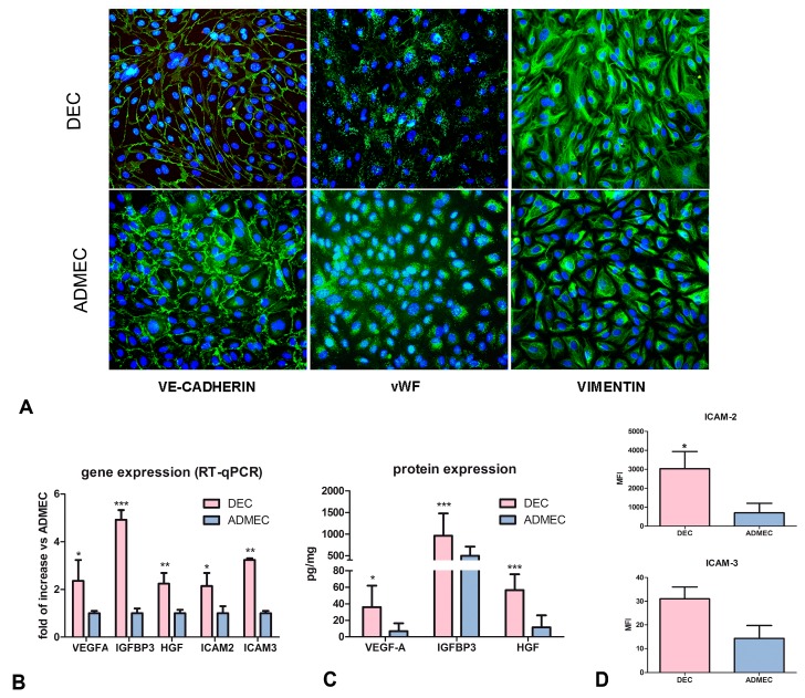
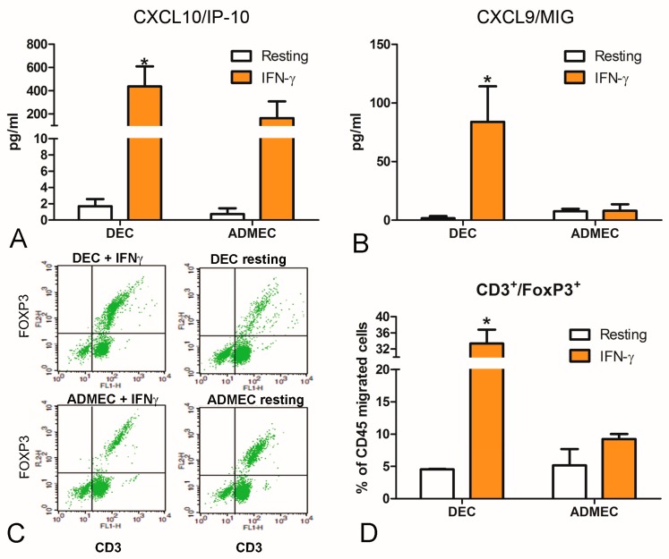
Results: DECs produced higher levels of HGF, VEGF-A and IGFBP3 compared to ADMECs. DECs expressed adhesion molecules, ICAM-2 and ICAM-3, and a mild response to TNF-α was observed. Finally, DECs produced high levels of CXCL9/MIG and CXCL10/IP-10 in response to IFN-γ and selectively recruited Treg lymphocytes.

Conclusion: DEC phenotype differs considerably from that of ADMECs, suggesting that DECs may play an active role in the control of immune response and angiogenesis at the foetal-maternal interface.

Keywords: angiogenesis; decidua; endothelium; inflammation; skin.


Conflict of interest statement

The authors declare that there is no conflict of interests regarding the publication of this article.

Figures

Figure 1
Figure 1
Phenotypic characterization of DECs and ADMECs. (A) Immunofluorescence analysis of vWF, VE-cadherin and vimentin on isolated and cultured DECs and ADMECs. Original magnification: 200×. (B) RT-qPCR of VEGF-A, HGF, IGFBP3, ICAM-2 and ICAM-3 genes differentially expressed by DECs and ADMECs. The data represent the mean ± SD of triplicate samples from five separate experiments, * p < 0.05, ** p < 0.01, *** p < 0.005. (C) Evaluation of the production of VEGF-A, HGF and IGFBP3 proteins in the supernatants of a confluent monolayer of DECs and ADMECs after 4 h of culture using a commercial ELISA kit. The data represent the mean ± SD of triplicate samples from five separate experiments. * p < 0.05, ** p < 0.01, *** p < 0.005. (D) Cytofluorimetric analysis for the expression of ICAM-2 and ICAM-3 in basal condition of freshly isolated DECs and ADMECs. The ECs were incubated with PE-conjugated mouse anti-human ICAM-2 and ICAM-3 mAb. PE-conjugated isotype-matched IgG2a or IgG1 were used as negative control, respectively. Data are represented as mean ± SD of the Mean Fluorescence Intensity (MFI) of five separate experiments.
Figure 2
Figure 2
Permeabilizing activity of endothelial cells to classical vasoactive stimuli. The permeabilizing activity was evaluated kinetically, after 5 (A), 15 (B) and 30 min (C) adding PAF (D), HIS (E) or BK (F), to the upper chamber of the TW, measuring the amount of FITC-labeled BSA that leaked through a monolayer of endothelial cells into the lower chamber. The data represent the mean ± SD of duplicate samples from four separate experiments * p < 0.05; ** p<0.01; *** p < 0.005.
Figure 3
Figure 3
Production of intracellular ROS by endothelial cells. ROS production by endothelial cells exposed to TNF-α (A) or HIS (B). Following pre-treatment with TNF-α (100 ng/mL) or histamine (HIS; 0.1 µM), ECs were stained with Ampliflu Red for the evaluation of H2O2 production after 30, 60 120, 180 and 240 min. (C) Histograms represent the production of intracellular ROS by endothelial cells stimulated with TNF-α or HIS after 120 min. The data represent the mean ± SD of triplicate samples from five separate experiments; * p < 0.05.
Figure 4
Figure 4
Secretion of chemokines and surface-expression of adhesion molecules by ECs stimulated with TNF-α. CXCL8/IL-8 (A), CCL2/MCP-1 (B), CCL3/MIP-1α (C) and CCL5/RANTES (D) production in DEC and ADMEC supernatants after 4 h incubation with TNF-α was measured using a beads-based multiplex immunoassay (Luminex®). (E) ELISA on the whole cells for the expression of ICAM-1, VCAM-1 or E-Selectin on DEC and ADMEC plasma membrane after 4 h or 18 h incubation with TNF-α. The data represent the mean ± SD of triplicate samples from five separate experiments * p < 0.01; ** p < 0.005. (F) Trans-endothelial migration of Lympho-Monocytes (LM) across untreated and TNF-α-treated DEC and ADMEC: migration is shown as the number of migrated cells. The data represent the mean ± SD of triplicate samples from three separate experiments * p < 0.05; n.s. not significant.
Figure 5
Figure 5
Secretion of chemokines and trans-endothelial migration of LM through EC stimulated with IFN-γ. The production of CXCL10/IP-10 (A) and CXCL9/MIG (B) in DEC and ADMEC supernatants after 4 h incubation with IFN-γ was measured using a beads-based multiplex immunoassay (Luminex®). The data represent the mean ± SD of triplicate samples from five separate experiments * p < 0.01. (C,D) Trans-endothelial migration of LM across untreated and IFN-γ-treated DEC and ADMEC. (C) Representative dot plots for flow cytometry of trans-endothelial migrated LM, stained for CD3 and FoxP3. (D) Quantitation of the percent of total migrated LM cells, positive for both CD3 and FoxP3 (CD3+FoxP3+) by flow cytometry. Migration was presented as the percentage of CD45 migrated cells. The data represent the mean ± SD of duplicate samples from three separate experiments * p < 0.05.

References

    1. Mantovani A., Bussolino F., Dejana E. Cytokine regulation of endothelial cell function. FASEB J. 1992;6:2591–2599. doi: 10.1096/fasebj.6.8.1592209. - DOI - PubMed
    1. Garlanda C., Dejana E. Heterogeneity of endothelial cells. Specific markers. Arterioscler. Thromb. Vasc. Biol. 1997;17:1193–1202. doi: 10.1161/01.ATV.17.7.1193. - DOI - PubMed
    1. Danese S., Dejana E., Fiocchi C. Immune regulation by microvascular endothelial cells: Directing innate and adaptive immunity, coagulation, and inflammation. J. Immunol. 2007;178:6017–6022. doi: 10.4049/jimmunol.178.10.6017. - DOI - PubMed
    1. Aird W.C. Endothelial cell heterogeneity. Cold Spring Harb. Perspect. Med. 2012;2:a006429. doi: 10.1101/cshperspect.a006429. - DOI - PMC - PubMed
    1. Robert C., Kupper T.S. Inflammatory skin diseases, T cells, and immune surveillance. N. Engl. J. Med. 1999;341:1817–1828. doi: 10.1056/NEJM199912093412407. - DOI - PubMed

MeSH terms