. 2019 Mar 31;8(4):298.
doi: 10.3390/cells8040298.

miR-1306 Mediates the Feedback Regulation of the TGF-β/SMAD Signaling Pathway in Granulosa Cells

Affiliations

miR-1306 Mediates the Feedback Regulation of the TGF-β/SMAD Signaling Pathway in Granulosa Cells

Liu Yang et al. Cells. .

Abstract

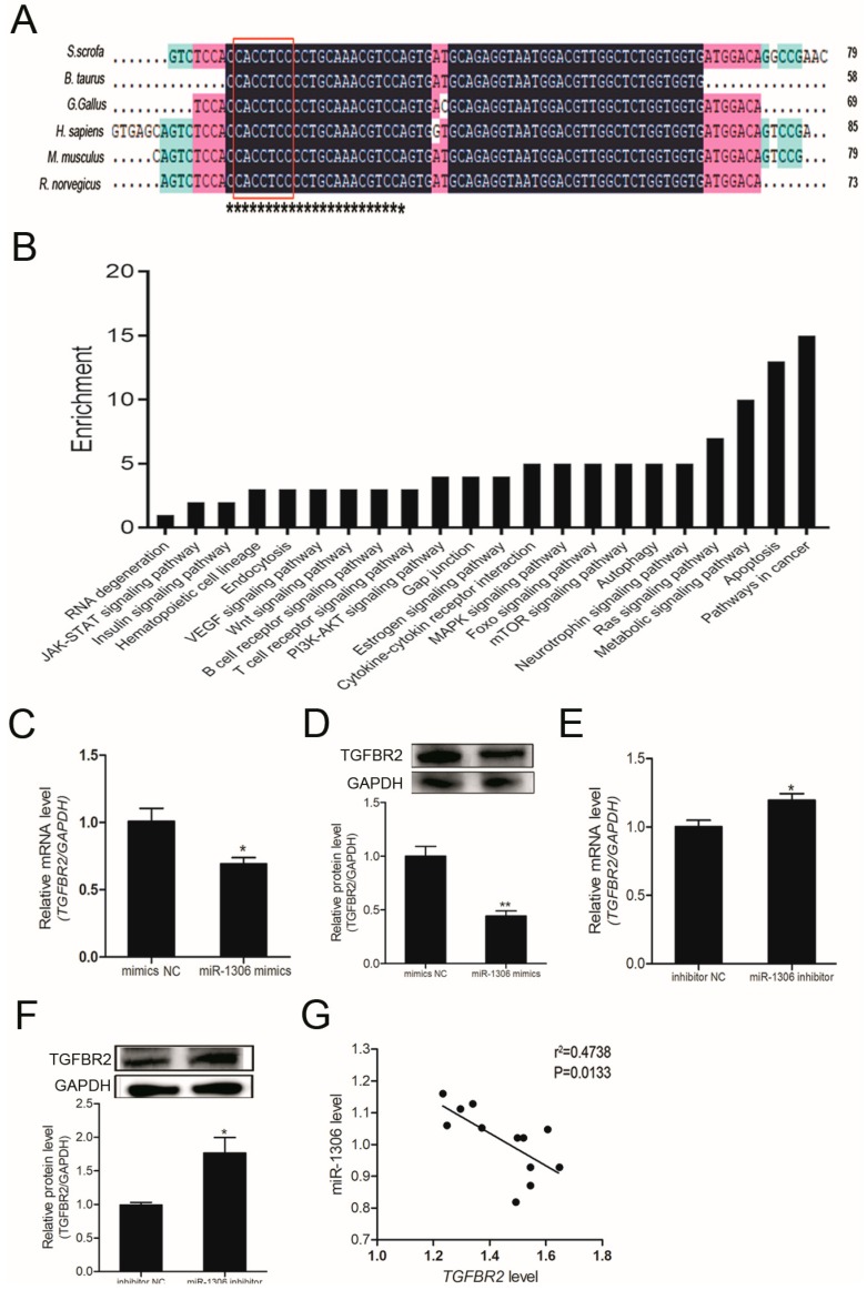
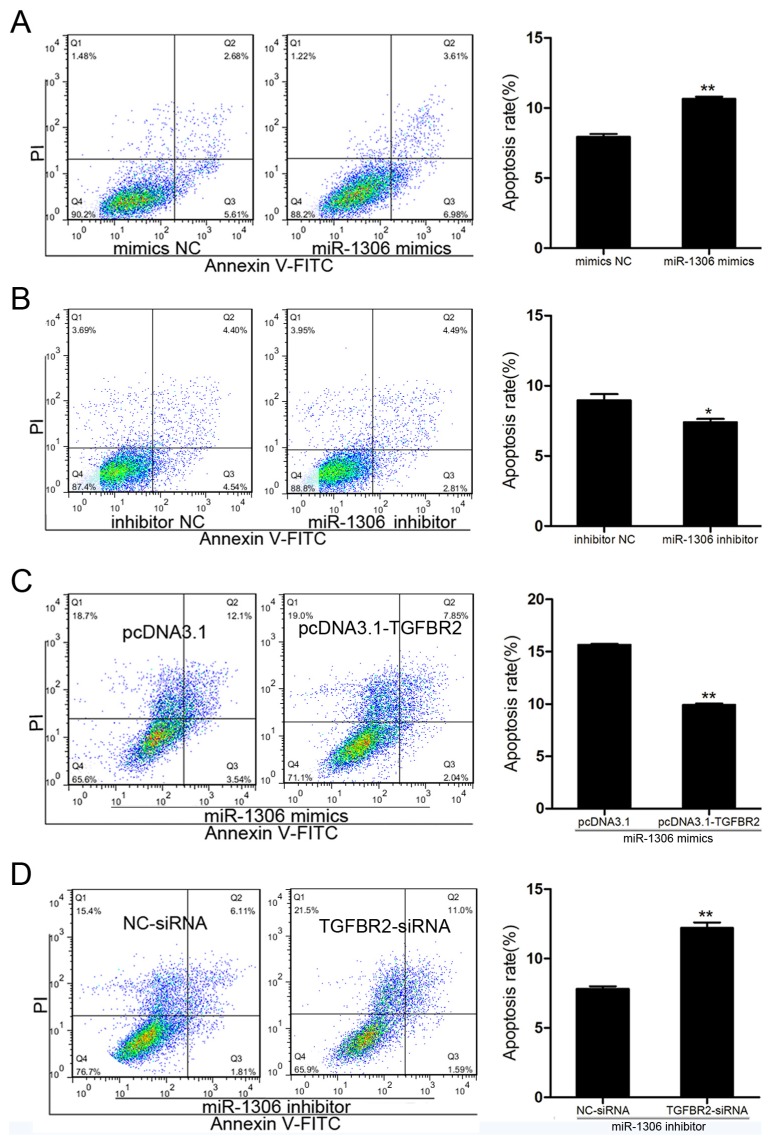
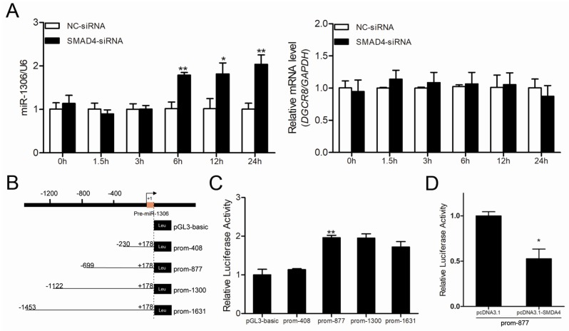
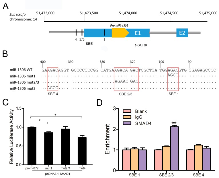
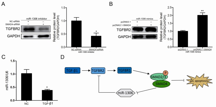
Transforming growth factor-β receptor II (TGFBR2), the type II receptor of the TGF-β/SMA- and MAD-related protein (SMAD) signaling pathway, plays a crucial role in TGF-β signal transduction and is regulated by multiple factors. Nevertheless, the modulation of the non-coding RNA involved in the process of TGFBR2 expression in ovaries is not well studied. In our study, we isolated and characterized the 3'-untranslated region (UTR) of the porcine TGFBR2 gene and microRNA-1306 (miR-1306) was identified as the functional miRNA that targets TGFBR2 in porcine granulosa cells (GCs). Functional analysis showed that miR-1306 promotes apoptosis of GCs as well as attenuating the TGF-β/SMAD signaling pathway targeting and impairing TGFBR2 in GCs. Moreover, we identified the miR-1306 core promoter and found three potential SMAD4-binding elements (SBEs). Luciferase and chromatin immunoprecipitation (ChIP) assays revealed that the transcription factor SMAD4 directly binds to the miR-1306 core promoter and inhibits its transcriptional activity. Furthermore, the TGF-β/SMAD signaling pathway is modulated by SMAD4 positive feedback via inhibition of miR-1306 expression in GCs. Collectively, our findings provide evidence of an epigenetic mechanism that modulates as well as mediates the feedback regulation of the classical TGF-β/SMAD signaling pathway in GCs from porcine ovaries.

Keywords: TGF-β/SMAD signaling pathway; TGFBR2; granulosa cell apoptosis; miR-1306.


Conflict of interest statement

The authors declare no conflict of interest.

Figures

Figure 1
Figure 1
MicroRNA-1306 (miR-1306) inhibits endogenous transforming growth factor-β receptor II (TGFBR2) expression in porcine granulosa cells (GCs). (A) Multiple-sequence alignment of pre-miR-1306 from six different species. miR-1306 mature sequences are indicated by black asterisks. Seed sequences of miR-1306 are indicated in red boxes. S. scrofa, Sus scrofa; B. Taurus, Bos taurus; G gorilla, Gorilla gorilla; H.sapiens, Homo sapiens; M.musculus, Mus musculus; R norvegicus, Rattus norvegicus. (B) Kyoto Encyclopedia of Genes and Genomes (KEGG) analysis showing the pathways targeted by miR-1306. (C,E) Results of Quantitative RT-PCR (qRT-PCR) showing TGFBR2 mRNA levels and (D,F) Western blotting results showing the TGFBR2 protein levels after miR-1306 mimics or inhibitor were transfected into porcine GCs, respectively (G) Correlation analysis between miR-1306 and TGFBR2 (n = 12). The data is represented by mean ± SEM. * indicates p < 0.05; ** indicates p < 0.01.
Figure 2
Figure 2
miR-1306 inactivates the transforming growth factor-β (TGF-β)/ SMA- and MAD-related protein (SMAD) signaling pathway in GCs by impairing TGFBR2. (A,B) Western blotting results showing levels of phospho-SMAD3 (p-SMAD3) and total-SMAD3 (t-SMAD3) protein after transfection of miR-1306 mimics or miR-1306 inhibitors into porcine GCs, respectively. (C,D) Western blotting results showing levels of p-SMAD3 and T-SMAD3 protein when both miR-1306 and TGFBR2 were overexpressed or silenced in porcine GCs. Each experiment has three independent repetition and results are shown as mean ± SEM. * indicates p < 0.05; ** indicates p < 0.01.
Figure 3
Figure 3
miR-1306 acts as an apoptotic factor in porcine GC through targeting TGFBR2. (A,B) Results showing the levels of GC apoptosis was (A) increased when cells were treated with mimic and (B) inhibited when cells were treated with miR-1306 inhibitor. The apoptotic cells were measured by fluorescence-activated cell sorting (FACS) (left panel) and the positive rates were calculated (right panels). (C,D) miR-1306 regulates GC apoptosis by inhibiting TGFBR2. (C) miR-1306 and TGFBR2 were both overexpressed or (D) silenced in porcine GCs. The apoptotic cells were identified by FACS (left panels) and the positive rates were calculated (right panel). Each experiment has three independent repetition and results are shown as mean ± SEM. * indicates p < 0.05; ** indicates p < 0.01.
Figure 4
Figure 4
SMAD4 suppresses miR-1306 expression by inhibiting its transcriptional activity independent of its host gene DGCR8. (A) The expression levels of miR-1306 (left) and its host gene DGCR8 (right) after SMAD4 silencing in porcine GCs were measured by qRT-PCR at different time intervals (0, 1.5, 3, 6, 12, 24 h). (B) Diagram depicting the 5′-flanking region of pre-miR-1306 and luciferase reporter vectors containing the candidate promoters of porcine miR-1306. (C) The recombinant vectors shown in panel B were transfected into porcine GCs and luciferase activities were detected. (D) miR-1306 promoter transcriptional activity in GCs treated with pcDNA™3.1 (pcDNA3.1)-SMAD4 was determined by Dual-Luciferase Activity Assay. Each experiment has three independent repetitions and results are shown as mean ± SEM. * indicates p < 0.05 and ** indicates p < 0.01.
Figure 5
Figure 5
SMAD4 acts as a transcription factor and directly binds to the miR-1306 promoter. (A) Schematic diagram depicting the genome locations of miR-1306 (yellow) and its host gene DGCR8 (blue), and the four potential SMAD4-binding sites are shown inside the black rectangles. (B) The luciferase reporter vectors containing porcine miR-1306 promoter with wild-type (prom-877) or mutant-type SBE motifs, shown in the red boxes. (C) pcDNA3.1-SMAD4 was co-transfected with recombinant vectors in (B) into GCs and luciferase activity assay was performed. (D) Chromatin immunoprecipitation (ChIP) assays and the enrichment were determined by qRT-PCR. Each experiment was independently repeated thrice and results are presented as mean ± SEM. * p means <0.05 and ** means p < 0.01.
Figure 6
Figure 6
miR-1306 mediates SMAD4 feedback regulation of the classical TGF-β/SMAD signaling pathway. Results of Western blot analysis showing the TGFBR2 protein level after (A) SMAD4- small interfering RNAs (SMAD4-siRNA) was transfected into miR-1306-inhibited GCs and after (B) pcDNA3.1-SMAD4 was transfected into miR-1306-overexpressed GCs. (C) miR-1306 expression levels in GCs were measured after TGF-β1 (20 ng/mL) treatment. (D) The SMAD4/miR-1306/TGFBR2 axis is involved in SMAD4-mediated positive feedback regulation in classical TGF-β/SMAD signaling pathway. Each experiment was independently repeated thrice and the results are presented as mean ± SEM. * p means <0.05 and ** means p < 0.01.

References

    1. Andl T., Le Bras G.F., Richards N.F., Allison G.L., Loomans H.A., Washington M.K., Andl C.D. Concerted loss of TGFβ-mediated proliferation control and E-cadherin disrupts epithelial homeostasis and causes oral squamous cell carcinoma. Carcinogenesis. 2014;35:2602–2610. doi: 10.1093/carcin/bgu194. - DOI - PMC - PubMed
    1. Mullen A.C., Wrana J.L. TGF-beta Family Signaling in Embryonic and Somatic Stem-Cell Renewal and Differentiation. Cold Spring Harb. Perspect. Biol. 2017;9 doi: 10.1101/cshperspect.a022186. - DOI - PMC - PubMed
    1. Massague J., Gomis R.R. The logic of TGFbeta signaling. FEBS Lett. 2006;580:2811–2820. doi: 10.1016/j.febslet.2006.04.033. - DOI - PubMed
    1. Heldin C.H., Landstrom M., Moustakas A. Mechanism of TGF-beta signaling to growth arrest, apoptosis, and epithelial-mesenchymal transition. Curr. Opin. Cell Biol. 2009;21:166–176. doi: 10.1016/j.ceb.2009.01.021. - DOI - PubMed
    1. Hill C.S. Transcriptional control by the SMADs. Cold Spring Harb. Perspect. Biol. 2016;8 doi: 10.1101/cshperspect.a022079. - DOI - PMC - PubMed

Publication types

MeSH terms

LinkOut - more resources