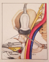
The relative risk to the femoral nerve as a function of patient positioning: potential implications for trigger point dry needling of the iliacus muscle
- PMID: 30935326
- PMCID: PMC6598541
- DOI: 10.1080/10669817.2019.1568699
The relative risk to the femoral nerve as a function of patient positioning: potential implications for trigger point dry needling of the iliacus muscle
Abstract
Objectives: Prudent dry needling techniques are commonly practiced with the intent to avoid large neurovascular structures, thereby minimizing potential excessive bleeding and neural injury. Patient position is one factor thought to affect the size of the safe zone during dry needling of some muscles. This study aimed to compare the size of the needle safe zone of the iliacus muscle during two different patient positions using ultrasound imaging. Methods: The distance from the anterior inferior iliac spine (AIIS) to the posterior pole of the femoral nerve was measured in 25 healthy participants (11 male, 14 female, mean age = 40) in both supine and sidelying positions using a Chison Eco1 musculoskeletal ultrasound unit. The average distance was calculated for each position and a two-tailed, paired t-test (α < 0.05) was used to examine the difference between positions. Results: The mean distance from the AIIS to the posterior pole of the femoral nerve was statistically greater with participants in the sidelying position (mean[SD] = 35.7 [6.2] mm) than in the supine position (mean[SD] = 32.1 [7.3] mm, p < .001). Discussion: Although more study is needed, these results suggest that patient positioning is one of several potential variables that should be considered in the optimization of patient safety/relative risk when performing trigger point dry needling. Level of Evidence: Level 4 (Pre-Post Test).
Keywords: Trigger point; dry needle; iliacus; patient position; risk; safety.
Figures


References
-
- APTA Description of dry needling in clinical practice: an educational resource paper. Alexandria, VA, USA: APTA Public Policy, Practice, and Professional Affairs Unit; 2013.
-
- Physiotherapy Acupuncture Association of New Zealand Guidelines for safe acupuncture and dry needling practice. Wellington, New Zealand: Physiotherapy Acupuncture Association of New Zealand; 2014.
-
- Mesa-Jimenez JA, Sanchez-Gutierrez J, de-la-Hoz-Aizpurua JL, et al. Cadaveric validation of dry needle placement in the lateral pterygoid muscle. Jmpt. 2015;38:145–150. - PubMed
-
- Hannah MC, Cope J, Palermo A, et al. Comparison of two angles of approach for trigger point dry needling of the lumbar multifidus in human donors (cadavers). Manual Ther. 2016;26:160–164. - PubMed
-
- Fernandez-de-Las Penas C, Mesa-Jimenez JA, Paredes-Mancilla JA, et al. Cadaveric and ultrasonographic validation of needling placement in the cervical multifidus muscle. JMPT. 2017;40:365–370. Dec;8(12):1168-1172. - PubMed
MeSH terms
LinkOut - more resources
Full Text Sources
Medical
