

The anatomical pathology of gout: a systematic literature review
- PMID: 30935368
- PMCID: PMC6444644
- DOI: 10.1186/s12891-019-2519-y
The anatomical pathology of gout: a systematic literature review
Abstract
Background: The aim of this systematic literature review was to comprehensively describe the anatomical pathology of tissues affected by gout.
Methods: We searched PubMed, The Cochrane Library, Excerpta Medica Database (EMBASE), and Web of Science Core Collection for all English language articles published before March 2018. Articles were included if they described the microscopic or macroscopic appearances of gout in human tissue.
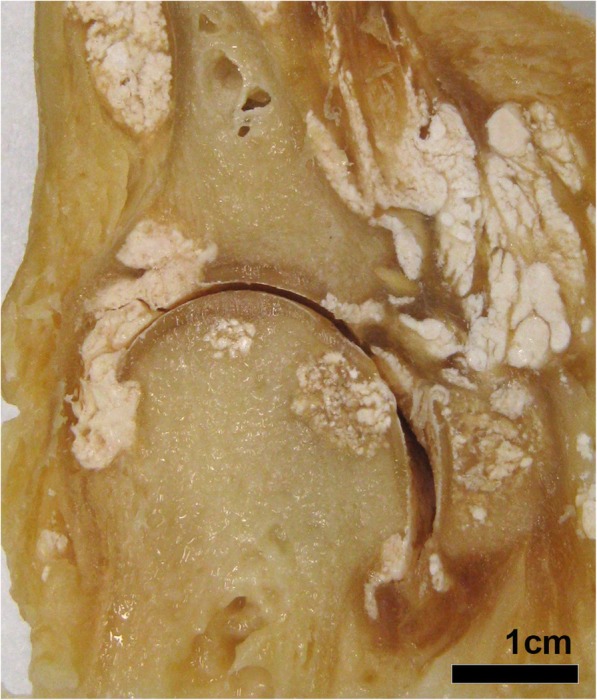
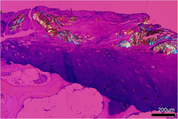
Results: Four hundred and seventeen articles met inclusion criteria and were included in the review. Articles describing the anatomical pathology of gout in musculoskeletal structures, including bone, tendon and ligaments, synovium and cartilage, were most common. Articles describing skin and kidney pathology in gout were also common, with pathology in other sites such as visceral organs less common. At all sites, monosodium urate crystal deposition was reported, and the tophus was also described within many different tissues. During a gout flare, diffuse acute neutrophilic synovial inflammation was evident. The tophus was described as an organised chronic giant cell granulomatous structure consisting of monosodium urate crystals, innate and adaptive immune cells, and fibrovascular tissue.
Conclusions: Consistent with the clinical presentation of gout, most studies describing the anatomical pathology of gout report involvement of musculoskeletal structures, with monosodium urate crystal deposition and tophus the most common lesions described. This review details the anatomical pathology features of gout at affected sites.
Keywords: Gout; Histology; Pathology; Synovium; Tophus.
Conflict of interest statement
Ethics approval and consent to participate
Human sample collection was approved by the Northern Regional ethics committee and all patients provided written informed consent. Collection and use of human cadaveric tissue was in accordance with the New Zealand Human Tissue Act 2008.
Consent for publication
Not applicable.
Competing interests
Dr. Dalbeth discloses the following: research grant funding from Amgen and AstraZeneca, speaker fees from Pfizer, Janssen, Horizon and Abbvie, consulting fees from Horizon and Kowa, outside the submitted work. The other authors declare that they have no competing interests.
Publisher’s Note
Springer Nature remains neutral with regard to jurisdictional claims in published maps and institutional affiliations.
Figures


References
-
- Taylor WJ, Fransen J, Jansen TL, Dalbeth N, Schumacher HR, Brown M, Louthrenoo W, Vazquez-Mellado J, Eliseev M, McCarthy G, et al. Study for updated gout classification criteria: identification of features to classify gout. Arthritis Care Res (Hoboken) 2015;67(9):1304–1315. doi: 10.1002/acr.22585. - DOI - PMC - PubMed
-
- Hench PS. Diagnosis and treatment of gout and gouty arthritis. J Am Med Assoc. 1941;116(6):453–455. doi: 10.1001/jama.1941.02820060001001. - DOI
Publication types
MeSH terms
Substances
LinkOut - more resources
Full Text Sources
Medical
Miscellaneous
