Optimal imaging conditions for the diagnosis of pleuroperitoneal communication
- PMID: 30936360
- PMCID: PMC6453368
- DOI: 10.1136/bcr-2018-228940
Optimal imaging conditions for the diagnosis of pleuroperitoneal communication
Abstract
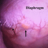
A 70-year-old woman with end-stage renal disease caused by a polycystic kidney disease developed massive right-sided pleural effusion 10 days after the initiation of peritoneal dialysis (PD). Although pleuroperitoneal communication (PPC) was suspected, computed tomographic peritoneography on usual breath holding did not show leakage. Therefore, we instructed her to strain with maximal breathing, which caused a jet of contrast material to stream from the peritoneal cavity into the right pleural cavity and allowed the identification of the exact site of the diaphragm defect. Following the thoracoscopic closure of the defect, she was discharged without recurrence of hydrothorax on PD. Hydrothorax due to PPC is a rare complication of PD. Notably, numerous previous modalities used to diagnose PPC lack sufficient sensitivity. Thus, an approach to spread the pressure gradient between the peritoneal cavity and the pleural cavity on imaging may improve this insufficient sensitivity.
Keywords: cardiothoracic surgery; chronic renal failure; dialysis; renal intervention.
© BMJ Publishing Group Limited 2019. Re-use permitted under CC BY-NC. No commercial re-use. See rights and permissions. Published by BMJ.
Conflict of interest statement
Competing interests: None declared.
Figures


References
-
- Fresenius Medical care. Fresenius Medical Care 2015 Annual Report. ESRD patients in 2015: A global prospective, 2015.
Publication types
MeSH terms
LinkOut - more resources
Full Text Sources
Medical
