. 2019 May;38(21):4182-4196.
doi: 10.1038/s41388-019-0797-3. Epub 2019 Apr 1.

Extracellular vesicle-dependent effect of RNA-binding protein IGF2BP1 on melanoma metastasis

Affiliations

Extracellular vesicle-dependent effect of RNA-binding protein IGF2BP1 on melanoma metastasis

Archita Ghoshal et al. Oncogene. 2019 May.

Abstract

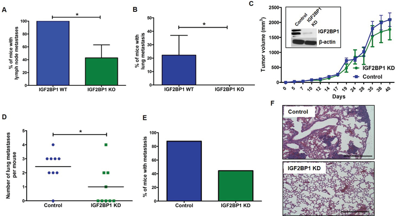
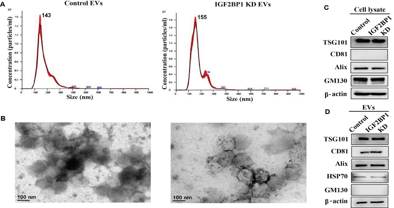
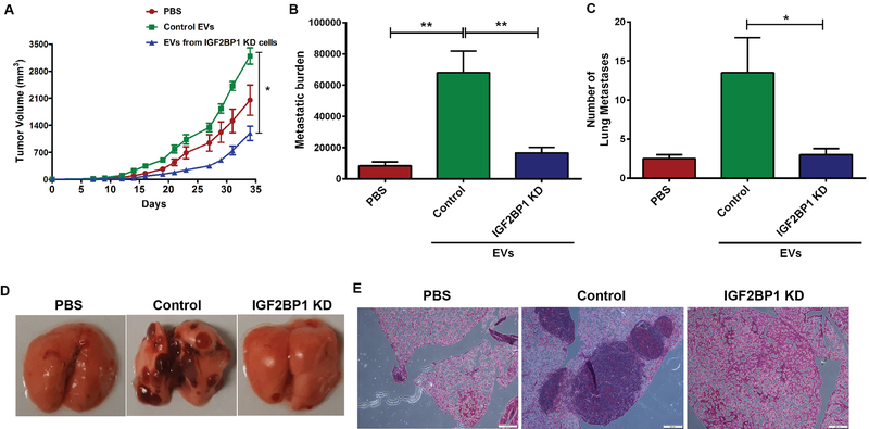
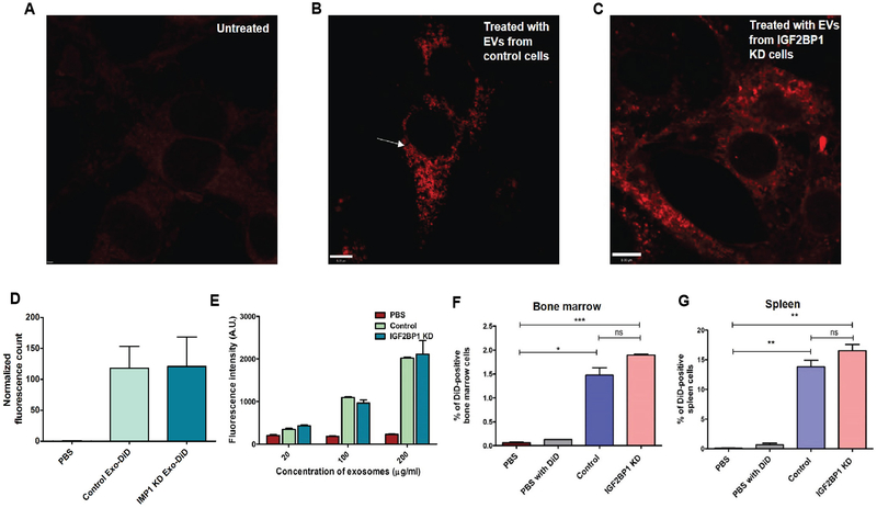
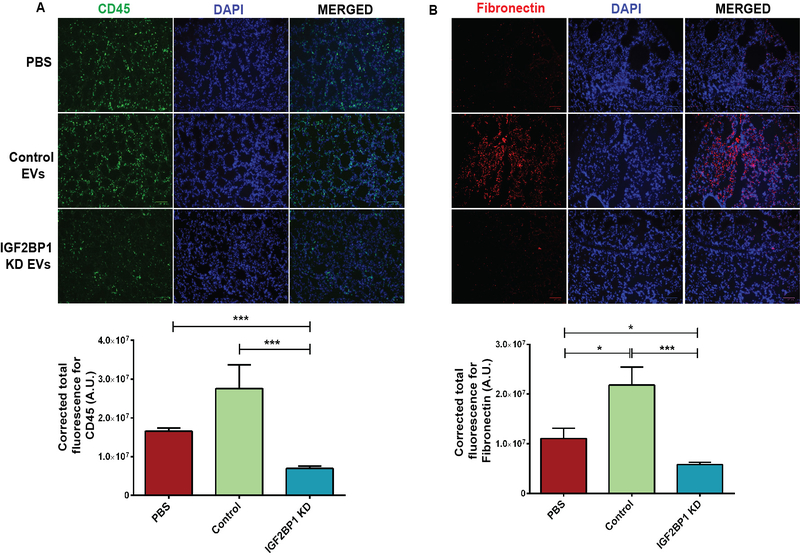
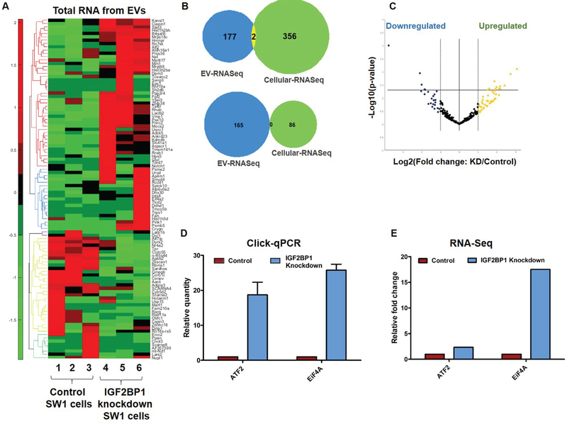
Insulin-like growth factor 2 mRNA-binding protein 1 (IGF2BP1) is a multifunctional RNA-binding protein with an oncofetal pattern of expression shown to be implicated in the development of a variety of malignancies. In this study, we explored the role and mechanisms of IGF2BP1 in melanoma development and progression. In two different in vivo models, we showed that although genetic deletion or shRNA-mediated suppression of IGF2BP1 did not affect primary tumor formation, it drastically suppressed lung metastasis. Here we demonstrated that extracellular vesicles (EVs) secreted by melanoma cells mediate the effects of IGF2BP1 on metastasis: EVs from the IGF2BP1 knockdown melanoma cells failed to promote metastasis, whereas EVs isolated from IGF2BP1-overexpressed melanoma cells further accelerated EV-induced metastasis. Moreover, the EVs from IGF2BP1 knockdown melanoma cells inhibited fibronectin deposition and accumulation of CD45+ cells in the lungs compared with control EVs, thus blocking the pre-metastatic niche formation potential of EVs. IGF2BP1 knockdown did not affect size, number, or protein/RNA concentration of secreted EVs or their uptake by recipient cells in vitro or in vivo. However, RNA-sequencing and proteomics analysis of the EVs revealed differential expression in a number of mRNA, proteins, and miRNAs. This suggested that IGF2BP1 is intimately involved in the regulation of the cargo of EVs, thereby affecting the pro-metastatic function of melanoma-derived EVs. To the best of our knowledge, this is the first study that demonstrates the role of RNA-binding protein IGF2BP1 in EV-mediated promotion of melanoma metastasis and may provide novel avenues for the development of metastatic inhibitors.


Conflict of interest statement

Disclosure of Potential Conflicts of Interest

The authors declare that there is no conflict of interest.

Figures

Figure 1.
Figure 1.
Metastasis study in two different mice models. A-B, Localized tamoxifen induction (4-HT 5mM) in IGF2BP1 –WT (Tyr::CreERT2 PTENloxP/loxP Braf F-V6000/+) and IGF2BP1 knockout (Tyr::CreERT2 PTENloxP/loxP Braf F-V6000/+ IGF2BP1loxP/loxP) mice. A, Percentage of mice with lymph node metastasis (p=0.0263), Student’s t-test. B, Percentage of mice with lung metastasis. IGF2BP1 WT mice had more lung metastasis compared to KO mice (p=0.0283), Student’s t-student. C-F, The role of IGF2BP1 was evaluated in vivo using a syngeneic metastasis model. The potential of lung metastasis of SW1 pInducer24 melanoma cell lines-untreated (control) and treated with doxycycline (to induce shRNA mediated knockdown of IGF2BP1) was evaluated in C3H mice. C, Primary tumor volume of mice belonging to control (n=8) and knockdown (KD) groups (n=10). Inset: Western blot showing knockdown of IGF2BP1 in mouse tissues. D, Number of lung metastasis developed in each mouse from the control and knockdown groups (p=0.0293), t-test. E, Percentage of lung metastasis in control mice compared to IGF2BP1 knockdown group. F, Microscopic evaluation of lungs of control and IGF2BP1 knockdown mice stained with H&E. Scale bar is 500 μm.
Figure 2.
Figure 2.
Analysis of EVs isolated from control and IGF2BP1 knockdown SW1 cells using sucrose cushion method. A, Nanoparticle tracking analysis (NTA). B, Transmission electron microscopy (TEM) imaging. C-D, Western blotting for starting cellular material and EVs to probe exosomal and golgi markers.
Figure 3.
Figure 3.
A, Analysis of primary tumor volume in C3H/HeJ mice injected with EVs (isolated with the sucrose cushion method) from control and IGF2BP1 knockdown cells over 35 days at an interval of 48 h; control group received PBS instead of EV injections. B, Metastatic burden in each group calculated by determining the area of metastatic lesions in H&E stained cross-sections of lungs using ImageJ software. C, Number of lung metastatic lesions in each group. D, Macroscopic imaging of lung metastasis in untreated mice and mice injected with either PBS or EVs from control or IGF2BP1 knockdown SW1 cells. E, Microscopic analysis of lung metastasis after H&E staining of sectioned lung tissues of mice injected with either PBS or EVs from control or IGF2BP1 knockdown SW1 cells.
Figure 4.
Figure 4.
Uptake of fluorescent DiD-labeled EVs isolated from control and IGF2BP1 knockdown cells. A, Fixed untreated control NIH3T3 cells viewed under STED confocal microscope. B-C, Fixed NIH3T3 cells viewed under STED confocal microscope to image uptake of DiD-labeled EVs following EV treatment for 24 h. B, Cells treated with control EVs, larger sized EVs are distinctly visible and marked with arrows. C, Cells treated with EVs from IGF2BP1 knockdown SW1 cells. D, Quantitative analyses of images acquired in STED microscope. E, Uptake of three different concentrations of EVs by recipient NIH3T3 cells. Fluorescence intensity of the cells was measured in a fluorescence spectrometer, after 24 hours of EV treatment. F, Graphical representation of flow cytometric analysis of bone marrow cells isolated from mice that received tail-vein injections of either PBS or DiD-PBS or DiD-labeled EVs isolated from control or IGF2BP1 knockdown SW1 cells, 24 hours prior to euthanization. G, Graphical representation of flow cytometric analysis of splenic cells isolated from the same mice.
Figure 5.
Figure 5.
Analysis of pre-metastatic niche formation in lungs. A, Expression of CD45 (top panel) with quantitative analysis in the bottom panel. B, Expression of fibronectin with quantitative analysis in the bottom panel. Expression of both CD45 and fibronectin increased markedly in the lungs of the mice treated with EVs (isolated with the sucrose cushion method) from control SW1 cells, as compared to the mice that did not receive EVs or the mice that received EVs from IGF2BP1 knockdown SW1 cells. Scale bar is 100 μm.
Figure 6.
Figure 6.
A, Heatmap analysis sequencing results of RNA from EVs. B, Venn diagrams depicting overlap of RNA sequenced from SW1 cells as well as EVs from SW1 cells, either control or IGF2BP1 knockdown. Top: upregulated genes, Bottom: downregulated genes. C, Volcano plot showing microRNA sequencing analysis from EVs. D, Real-time PCR analysis to probe expressions of ATF2 and EiF4A2 genes in EU-labeled RNA from NIH3T3 cells treated with EVs derived from EU-labeled control and IGF2BP1 knockdown SW1 cells. E, Graphical representation of the relative fold change of the same genes in the EVs as seen in the sequencing analysis of EVs.

References

    1. Dimitriadis E, Trangas T, Milatos S, Foukas PG, Gioulbasanis I, Courtis N, et al. Expression of oncofetal RNA-binding protein CRD-BP/IMP1 predicts clinical outcome in colon cancer. Int J Cancer. 2007;121(3):486–94. - PubMed
    1. Bell JL, Turlapati R, Liu T, Schulte JH, Huttelmaier S. IGF2BP1 harbors prognostic significance by gene gain and diverse expression in neuroblastoma. J Clin Oncol. 2015;33(11):1285–93. - PubMed
    1. Vikesaa J, Hansen TV, Jonson L, Borup R, Wewer UM, Christiansen J, et al. RNA-binding IMPs promote cell adhesion and invadopodia formation. EMBO J. 2006;25(7):1456–68. - PMC - PubMed
    1. Elcheva I, Goswami S, Noubissi FK, Spiegelman VS. CRD-BP protects the coding region of betaTrCP1 mRNA from miR-183-mediated degradation. Mol Cell. 2009;35(2):240–6. - PMC - PubMed
    1. Goswami S, Tarapore RS, Poenitzsch Strong AM, TeSlaa JJ, Grinblat Y, Setaluri V, et al. MicroRNA-340-mediated degradation of microphthalmia-associated transcription factor (MITF) mRNA is inhibited by coding region determinant-binding protein (CRD-BP). The Journal of biological chemistry. 2015;290(1):384–95. - PMC - PubMed

Publication types