. 2019 Apr;26(4):315-321.
doi: 10.1038/s41594-019-0208-z. Epub 2019 Apr 1.

Broad-spectrum enzymatic inhibition of CRISPR-Cas12a

Affiliations

Broad-spectrum enzymatic inhibition of CRISPR-Cas12a

Gavin J Knott et al. Nat Struct Mol Biol. 2019 Apr.

Abstract

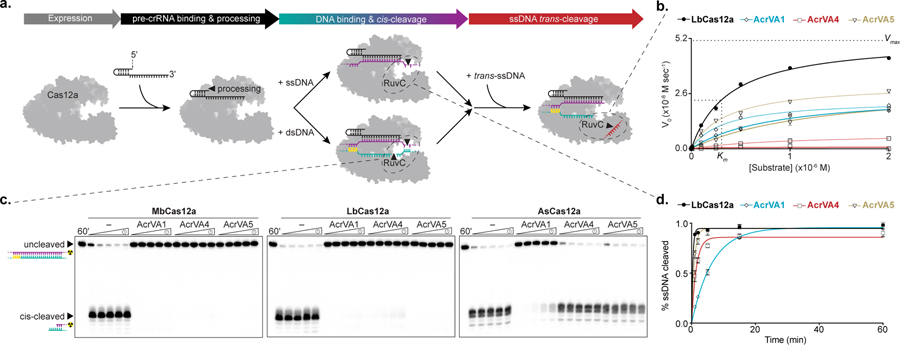
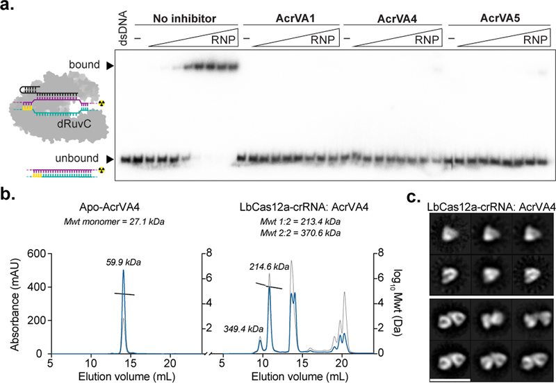
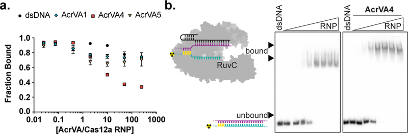
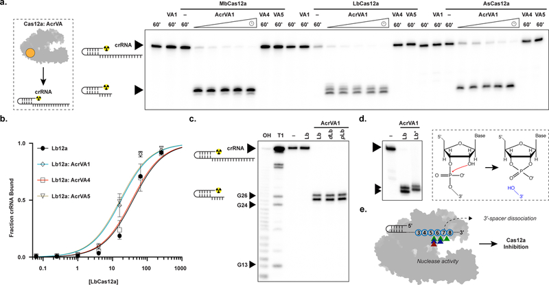
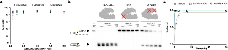
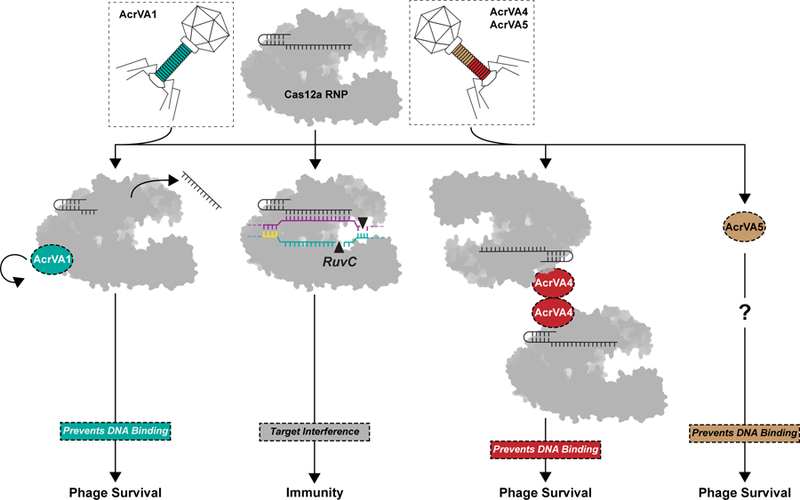
Cas12a is a bacterial RNA-guided nuclease used widely for genome editing and, more recently, as a molecular diagnostic. In bacteria, Cas12a enzymes can be inhibited by bacteriophage-derived proteins, anti-CRISPRs (Acrs), to thwart clustered regularly interspaced short palindromic repeat (CRISPR) adaptive immune systems. How these inhibitors disable Cas12a by preventing programmed DNA cleavage is unknown. We show that three such inhibitors (AcrVA1, AcrVA4 and AcrVA5) block Cas12a activity via functionally distinct mechanisms, including a previously unobserved enzymatic strategy. AcrVA4 and AcrVA5 inhibit recognition of double-stranded DNA (dsDNA), with AcrVA4 driving dimerization of Cas12a. In contrast, AcrVA1 is a multiple-turnover inhibitor that triggers cleavage of the target-recognition sequence of the Cas12a-bound guide RNA to irreversibly inactivate the Cas12a complex. These distinct mechanisms equip bacteriophages with tools to evade CRISPR-Cas12a and support biotechnological applications for which multiple-turnover enzymatic inhibition of Cas12a is desirable.


Conflict of interest statement

COMPETING INTERESTS

The Regents of the University of California have patents pending for CRISPR technologies on which the authors are inventors. J.A.D. is a co-founder of Caribou Biosciences, Editas Medicine, Intellia Therapeutics, Scribe Therapeutics, and Mammoth Biosciences. J.A.D. is a scientific advisory board member of Caribous Biosciences, Intellia Therapeutics, eFFECTOR Therapeutics, Scribe Therapeutics, Synthego, Metagenomi, Mammoth Biosciences, and Inari. J.A.D. is a Director at Johnson & Johnson and has sponsored research projects by Pfizer, Roche Biopharma, and Biogen.

Figures

Figure 1 –
Figure 1 –. AcrVAs do not inhibit all modes of DNA targeting by Cas12a.
a) Schematic representation of the steps in Cas12a target interference, b) Michaelis-Menten fit for 0.1 nM effective LbCas12a holoenzyme in the absence (black) or presence of AcrVA1 (blue), AcrVA4 (red), or AcrVA5 (yellow). The mean initial velocity (V0) is plotted against increasing DNase-Alert substrate concentrations (µM), where n = 3 replicates. The Vmax and Km for wild-type LbCas12a RuvC are indicated with dashed lines. c) Radiolabeled kinetic dsDNA cleavage assays for (left to right) MbCas12a, LbCas12a, and AsCas12a complexes with or without AcrVAs. Time courses represent 1’, 2’, 5’, 15’, and 60’. The uncleaved and cis-cleaved fractions are indicated with black triangles, d) Quantified percentage ssDNA cleaved for LbCas12a in the presence or absence of AcrVAs (mean ± sd, n = 3 independent experiments). Experimental fits are shown as solid lines and the calculated pseudo-first-order rate constants (kobs) (mean ± sd) are 2.6 ± 0.3 min−1, 0.15 ± 0.01 min−1, 0.7 ± 0.06 min−1, and 1.2 ± 0.09 min−1 for LbCas12a, LbCas12a+AcrVA1, LbCas12a+AcrVA4, and LbCas12a+AcrVA5 respectively. Source data for panels b and d are available with the paper online. The uncropped gel images are available in Supplementary Data Set 1.
Figure 2 –
Figure 2 –. AcrVAs block dsDNA binding and AcrVA4 dimerizes Cas12a.
a) Radiolabeled dsDNA electrophoretic mobility shift assay of increasing concentrations of dLbCas12a-crRNA complexed with or without AcrVA before association with dsDNA. The bound and unbound fractions are indicated with black triangles, b) Size exclusion chromatography coupled light scattering traces for (left) AcrVA4 alone and (right) LbCas12a-crRNA complexed with AcrVA4. The absorbance at 280 nm (blue) and 260 nm (grey) are shown (left axis) with the linear region for the mass estimate corresponding to the relevant peaks (black lines, central and right axis). The predicted molecular weights for each sample are shown above the graph and the calculated molecular weights are indicated adjacent to the relevant peak c) 2D-class averages of LbCas12a-crRNA monomers (top) and LbCas12a-crRNA dimers bound to AcrVA4 (bottom). The scale bar represents 28 nm. The uncropped gel images are available in Supplementary Data Set 1.
Figure 3 –
Figure 3 –. AcrVA4 displaces dsDNA bound to dCas12a but not wild-type Cas12a.
a) Quantified fraction dsDNA bound by dLbCas12a-crRNA after complexing with dsDNA before the addition of increasing concentrations of AcrVA or dsDNA competitor determined by EMSA (mean ± sd, n = 3 independent experiments), Source data are available with the paper online. b) Radiolabeled dsDNA electrophoretic mobility shift assay of increasing concentrations of LbCas12a-crRNA first complexed with dsDNA before addition of AcrVA. The bound and unbound fractions are indicated with black triangles. The uncropped gel images are available in Supplementary Data Set 1.
Figure 4 –
Figure 4 –. AcrVA1-triggered endoribonuclease activity truncates a Cas12a-bound crRNA.
a) Radiolabeled kinetic crRNA cleavage assays for (left to right) MbCas12a, LbCas12a, and AsCas12a complexed with or without AcrVAs. Time courses represent 1, 2, 5, 15, and 60 min. Black triangles indicate full-length and truncated crRNA, b) Quantified fraction crRNA bound by LbCas12a in the presence or absence of AcrVAs determined by EMSA (mean ± sd, n = 3 independent experiments). Measured dissociation constants (Kd) are 38.9 nM ± 4.7, 17.6 ± 2.4, 35.8 ± 4.4, and 16.4 ± 2.1 in absence of inhibitor or in the presence of AcrVA1, AcrVA4, or AcrVA5, respectively, Source data are available with the paper online. c) Radiolabeled crRNA cleavage assay with LbCas12a-crRNA complexed without or with AcrVA1. Treatments in the absence of AcrVA1 are (left to right) crRNA hydrolysis ladder (OH), crRNA RNase T1 digestion (T1), untreated crRNA (−), and crRNA incubated with LbCas12a (Lb). Treatments in the presence of AcrVA1 are (left to right) wild-type LbCas12a (Lb), dLbCas12a (D832A, dLb), and processing dead Cas12a (K785A, pLb). A large black triangle indicates the full-length crRNA, smaller triangles indicate RNase T1 mapped G-nucleotides, d) Radiolabeled crRNA cleavage assay using LbCas12a-crRNA complexed with AcrVA1 that is either untreated (Lb) or treated with PNK (Lb*), e) Schematic representation of AcrVA1-triggered crRNA spacer cleavage activity on Cas12a. Cleavage sites for Mb (blue), Lb (green), and AsCas12a (red) are indicated with triangles. The uncropped gel images are available in Supplementary Data Set 1.
Figure 5 –
Figure 5 –. AcrVA1-triggered endoribonuclease activity is multiple turnover, requires the PID, and competes with AcrVA5.
a) Percentage crRNA spacers truncated after 1 hr at different ratios of AcrVA1:Cas12a-crRNA (mean ± sd, n = 3 independent experiments). b) Radiolabeled kinetic crRNA cleavage assays for (left to right) LbCas12a, ∆PID LbCas12a, and ∆REC1/2 LbCas12a in the presence of AcrVA1. Time courses represent 1, 2, 5, 15, and 60 min. Black triangles indicate the full-length and truncated crRNA. The approximate position of the truncated domains is shown with a red cross. c) Quantified time-course of percentage AcrVA1-triggered spacers truncated in the presence AcrVA4 or AcrVA5 (n = 3 independent experiments). Experimental fits are shown as solid lines and the calculated pseudo-first-order rate constants (kobs) (mean ± sd) are 4.8 ± 0.2 min−1, 4.4 ± 0.3 min−1, and 0.5 ± 0.04 min−1 for AcrVA1, AcrVA1 + AcrVA4, and AcrVA1 + AcrVA5, respectively. Source data for panels a and c are available with the paper online. The uncropped gel images are available in Supplementary Data Set 1.
Figure 6 –
Figure 6 –. Three distinct modes of CRISPR-Cas12a inactivation.
Model for AcrVA1, AcrVA4, and AcrVA5 inhibition of Cas12a. Cas12a assembles with its crRNA to form a surveillance complex (top). In the absence of inhibitors, Cas12a recognizes a complementary target DNA activating the RuvC leading to target interference and immunity. Phage-encoded AcrVA1 (teal) associates with Cas12a triggering crRNA spacer truncation preventing DNA binding. Phage-encoded AcrVA4 dimerizes (red) Cas12a and blocks dsDNA binding. Phage-encoded AcrVA5 (brown) blocks Cas12a dsDNA binding via an unknown mechanism.

References

    1. Wright AV, Nuñez JK & Doudna JA Biology and Applications of CRISPR Systems: Harnessing Nature’s Toolbox for Genome Engineering. Cell 164, 29–44 (2016). - PubMed
    1. Brouns SJJ et al. Small CRISPR RNAs Guide Antiviral Defense in Prokaryotes. Science (80-. ) 321, 960–964 (2008). - PMC - PubMed
    1. Marraffini LA & Sontheimer EJ CRISPR interference limits horizontal gene transfer in staphylococci by targeting DNA. Science (80-. ) 322, 1843–1845 (2008). - PMC - PubMed
    1. Barrangou R et al. CRISPR provides acquired resistance against viruses in prokaryotes. Science (80-. ) 315, 1709–1712 (2007). - PubMed
    1. Garneau JE et al. The CRISPR/Cas bacterial immune system cleaves bacteriophage and plasmid DNA. Nature 468, 67–71 (2010). - PubMed

Publication types

MeSH terms

Substances