

Chronic eosinophilic pneumonia: clinical perspectives
- PMID: 30936702
- PMCID: PMC6420789
- DOI: 10.2147/TCRM.S157882
Chronic eosinophilic pneumonia: clinical perspectives
Abstract
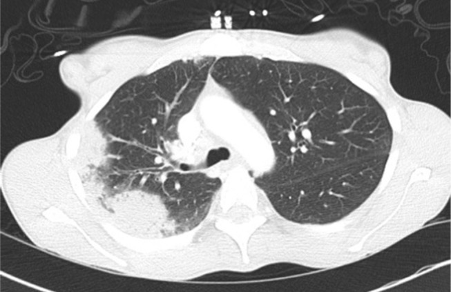
Chronic eosinophilic pneumonia (CEP) is an eosinophilic lung disease that is typically diagnosed by a triad of clinical symptoms including pulmonary symptoms, eosinophilia and characteristic radiographic abnormalities. It requires a high index of suspicion given its overlap with other eosinophilic conditions and lack of a specific diagnostic test. The diagnosis is made after careful consideration of other secondary causes of eosinophilia, such as infectious, drugs, or toxic etiologies. CEP generally responds rapidly to treatment, which primarily consists of corticosteroid therapy, but relapses are common. Novel therapies are being explored as more information is being discovered about the pathophysiology of eosinophilic disease processes. Close follow-up is important given the difficulty in weaning patients from glucocorticoids with many patients developing sequelae of chronic glucocorticoid therapy. Therefore, exploring alternative treatments is of upmost importance.
Keywords: BAL; IL-5; eosinophilic; infiltrates; lung; mepolizumab; prednisone.
Conflict of interest statement
Disclosure The authors report no conflicts of interest in this work.
Figures


References
-
- Cottin V, Cordier JF. Eosinophilic lung diseases. Immunol Allergy Clin North Am. 2012;32(4):557–586. - PubMed
-
- Rose DM, Hrncir DE. Primary eosinophilic lung diseases. Allergy Asthma Proc. 2013;34(1):19–25. - PubMed
-
- Carrington CB, Addington WW, Goff AM, et al. Chronic eosinophilic pneumonia. N Engl J Med. 1969;280(15):787–798. - PubMed
-
- Cottin V. Diseases Elung. Eosinophilic lung diseases. Clin Chest Med. 2016;37(3):535–556. - PubMed
Publication types
LinkOut - more resources
Full Text Sources
