. 2019 Mar 14:11:2139-2150.
doi: 10.2147/CMAR.S188648. eCollection 2019.

Oncogenic role of ABHD5 in endometrial cancer

Affiliations

Oncogenic role of ABHD5 in endometrial cancer

Qing Zhou et al. Cancer Manag Res. .

Abstract

Background: Abhydrolase domain containing 5 (ABHD5) functions as a tumor suppressor in colorectal and prostate cancers. The aim of this study was to investigate the roles of ABHD5 in endometrial cancer.

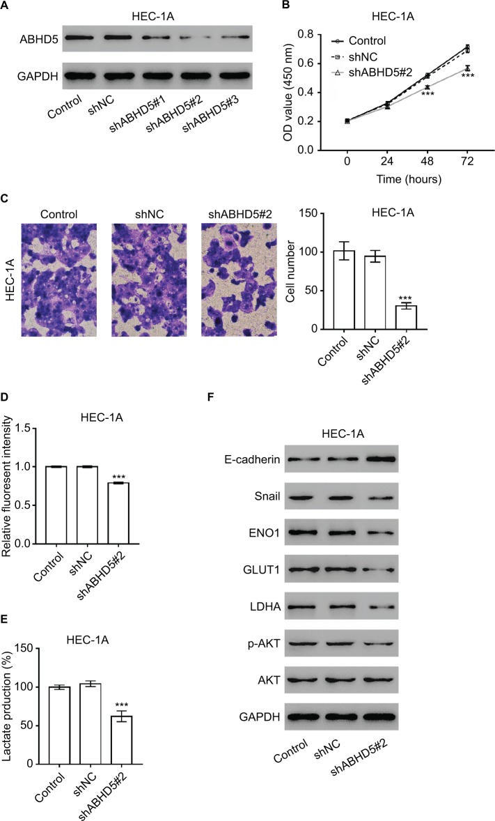
Materials and methods: ABHD5 expression was detected in clinical samples by immunohistochemical staining. Cell proliferation and invasion were evaluated with the Cell Counting Kit-8 and Transwell assay, respectively. Western blotting was performed to analyze protein expression. Glucose uptake was assessed by 2-[N-(7-nitrobenz-2-oxa-1,3-diazol-4-yl)amino]-2-deoxyglucose. Lactate production was detected by a lactate assay kit.

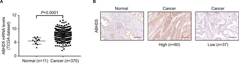
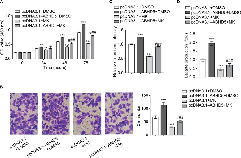
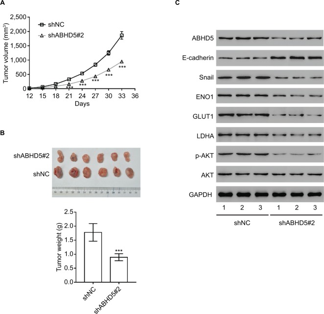
Results: In the present study, ABHD5 was overexpressed in endometrial cancer tissues, and its expression was closely correlated with the International Federation of Gynecology and Obstetrics (FIGO) stage and lymph node metastasis. In addition, we observed that the knockdown of ABHD5 inhibited cell proliferation, invasion, glucose uptake and lactate production in HEC-1A cells, which expressed high levels of ABHD5. Conversely, the opposite effects were observed when ABHD5 was ectopically expressed in Ishikawa cells, which had low levels of ABHD5. Furthermore, the changes in glycolysis regulators (enolase 1 [ENO1], glucose transporter 1 [GLUT1] and lactate dehydrogenase A [LDHA]) and epithelial-to-mesenchymal transition-related proteins (E-cadherin and Snail) in HEC-1A cells with ABHD5 knockdown were consistent with the effects of ABHD5 on glycolysis and cell invasion. Phosphatase and tensin homolog deleted on chromosome 10 (PTEN) was increased, while the phosphorylated AKT (p-AKT) was decreased when ABHD5 was downregulated. Notably, treatment with the allosteric AKT inhibitor MK-2206 completely abolished the effects caused by ABHD5 overexpression in Ishikawa cells. Finally, ABHD5 knockdown potently suppressed tumor growth in vivo.

Conclusion: Overall, these results suggest that ABHD5 may play an oncogenic role in endometrial cancer via the AKT pathway.

Keywords: ABHD5; AKT; endometrial cancer; glycolysis.


Conflict of interest statement

Disclosure The authors report no conflicts of interest in this work.

Figures

Figure 1
Figure 1
ABHD5 overexpression in endometrial cancer. Notes: (A) The expression level of ABHD5 in endometrial cancer and normal tissues based on TCGA dataset. (B) Expression of ABHD5 was determined in endometrial cancer tissues and normal tissues by immunohistochemistry staining. Scale bar: 100 µm. Abbreviation: TCGA, The Cancer Genome Atlas.
Figure 2
Figure 2
ABHD5 knockdown suppressed endometrial cancer cell proliferation, invasion and the Warburg effect. Notes: (A) HEC-1A cells were transduced with ABHD5 shRNA and control shRNA (shNC). Cells without any treatment served as a control. Western blot analysis was performed to evaluate the efficiency of three ABHD5 shRNAs in HEC-1A cells at 48 hours post treatment. Representative blots from three independent experiments are shown. (B) Cell proliferation was determined by CCK-8 assay (n=3 biological replicates) in HEC-1A cells transfected with shABHD5#2 and shNC. (C) A Transwell assay (n=3 biological replicates) was performed to assess cell invasive ability, magnification 200×. (D) 2-NBDG uptake was measured (n=3 biological replicates) at 24 hours post treatment. Fluorescent intensity was normalized to protein content and then divided by the value of the control. (E) Lactate production (n=3 biological replicates) was measured and divided by the value of the control. (F) Western blot analysis was performed to detect the related proteins. Representative blots from three independent experiments are shown. ***P<0.001 vs the control and shNC. Abbreviations: 2-NBDG, 2-[N-(7-nitrobenz-2-oxa-1,3-diazol-4-yl) amino]-2-deoxyglucose; CCK-8, Cell Counting Kit-8.
Figure 3
Figure 3
The activation of AKT is necessary for the biological functions of ABHD5 in endometrial cancer cells. Notes: Ishikawa cells were transfected with pcDNA3.1–ABHD5 or pcDNA3.1 and treated with 10 µM MK-2206 (MK; Merck Millipore, Billerica, MA, USA) or vehicle (DMSO). (A) Cell proliferation was detected by CCK-8 assay (n=3 biological replicates). (B) Cell invasion was detected by Transwell assay (n=3 biological replicates), magnification 200×. (C) 2-NBDG uptake was measured (n=3 biological replicates). Fluorescent intensity was normalized to protein content and then divided by the value of the control (the first column). (D) Lactate production (n=3 biological replicates) was measured and divided by the value of the control (the first column). **P<0.01, ***P<0.001 vs pcDNA3.1+DMSO; #P<0.05, ###P<0.001 vs pcDNA3.1+MK. Abbreviations: 2-NBDG, 2-[N-(7-nitrobenz-2-oxa-1,3-diazol-4-yl) amino]-2-deoxyglucose; CCK-8, Cell Counting Kit-8; DMSO, dimethylsulfoxide; MK, MK-2206.
Figure 4
Figure 4
Impact of ABHD5 knockdown on endometrial cancer growth in vivo. Notes: (A, B) ABHD5 knockdown significantly blocked xenograft growth in nude mice (n=6 per group). Tumor volume was measured every 3 days. On day 33 after inoculation, the mice were sacrificed and tumors were weighed (n=6). (C) Western blotting was performed to assess the expression of the indicated proteins in xenografts. Representative blots from three independent experiments are shown. ***P<0.001.

References

    1. Ali AT. Risk factors for endometrial cancer. Ceska Gynekol. 2013;78(5):448–459. - PubMed
    1. Vander Heiden MG, Cantley LC, Thompson CB. Understanding the Warburg effect: the metabolic requirements of cell proliferation. Science. 2009;324(5930):1029–1033. - PMC - PubMed
    1. Koppenol WH, Bounds PL, Dang CV. Otto Warburg’s contributions to current concepts of cancer metabolism. Nat Rev Cancer. 2011;11(5):325–337. - PubMed
    1. Bayley JP, Devilee P. The Warburg effect in 2012. Curr Opin Oncol. 2012;24(1):62–67. - PubMed
    1. Dang CV, Le A, Gao P. Myc-induced cancer cell energy metabolism and therapeutic opportunities. Clin Cancer Res. 2009;15(21):6479–6483. - PMC - PubMed