. 2019 Feb 22:2:2515256418825409.
doi: 10.1177/2515256418825409.

The Endoplasmic Reticulum-Mitochondria Encounter Structure Complex Coordinates Coenzyme Q Biosynthesis

Affiliations

The Endoplasmic Reticulum-Mitochondria Encounter Structure Complex Coordinates Coenzyme Q Biosynthesis

Michal Eisenberg-Bord et al. Contact (Thousand Oaks). .

Abstract

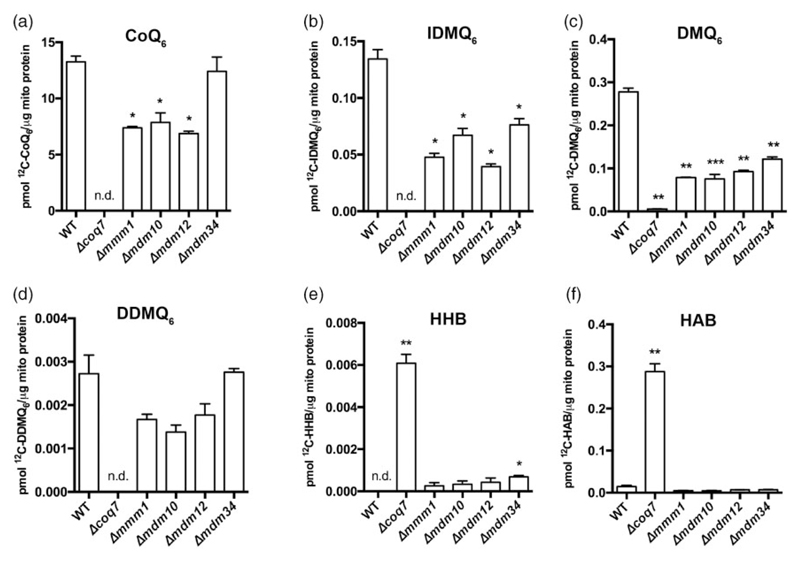
Loss of the endoplasmic reticulum (ER)-mitochondria encounter structure (ERMES) complex that resides in contact sites between the yeast ER and mitochondria leads to impaired respiration; however, the reason for that is not clear. We find that in ERMES null mutants, there is an increase in the level of mRNAs encoding for biosynthetic enzymes of coenzyme Q6 (CoQ6), an essential electron carrier of the mitochondrial respiratory chain. We show that the mega complexes involved in CoQ6 biosynthesis (CoQ synthomes) are destabilized in ERMES mutants. This, in turn, affects the level and distribution of CoQ6 within the cell, resulting in reduced mitochondrial CoQ6. We suggest that these outcomes contribute to the reduced respiration observed in ERMES mutants. Fluorescence microscopy experiments demonstrate close proximity between the CoQ synthome and ERMES, suggesting a spatial coordination. The involvement of the ER-mitochondria contact site in regulation of CoQ6 biogenesis highlights an additional level of communication between these two organelles.

Keywords: ER-mitochondrial encounter structure; coenzyme Q; endoplasmic reticulum; mitochondrion (mitochondria).


Conflict of interest statement

Declaration of Conflicting Interests The author(s) declared no potential conflicts of interest with respect to the research, authorship, and/or publication of this article.

Figures

Figure 1
Figure 1. Cells lacking ERMES show higher levels of COQ mRNAs without alterations to Coq proteins.
(a) Levels of mRNAs of the indicated COQ genes were measured in different W303-based strains deleted for ERMES subunits (Δmmm1, Δmdm10, Δmdm12 Δmdm34), and a control strain, during growth in medium containing glycerol. The majority of COQ biosynthetic genes show higher mRNA levels compared to the control. Values are averages of two biological repeats. (b) Immunoblotting for steady-state levels of Coq polypeptides in purified mitochondria isolated from the indicated W303-based strains. Shown are mutants of the ERMES complex (Δmmm1, Δmdm10, Δmdm12, and Δmdm34), Δcoqcoq3coq10) and a control, demonstrating that steady-state levels of the different Coq polypeptides were not dramatically altered in the ERMES deletion strains. Immunoblotting was performed with antisera against designated yeast Coq polypeptides (Coq3-Coq10), Mdh1 as a mitochondrial marker, and Sec62 as an ER marker. Arrows denote the corresponding protein in their respective blots. Images are representative gels from at least two biological replicates.
Figure 2
Figure 2. The CoQ synthome is destabilized in the absence of ERMES subunits.
(a) Two-dimensional Blue Native-SDS-PAGE gel electrophoresis analysis of the CoQ synthome in mitochondria from ERMES mutants or from a wild-type (WT) control strain. Gels were immunoblotted against Coq4. In the control sample, the CoQ synthome appears as complexes ranging from 232 kDa to >1 MDa (the positions of the synthome is marked by the yellow arrow), while in the ΔERMES strains, the high molecular weight signal is replaced by a signal dispersed over a range of smaller molecular weights (indicated by brackets). The relevant band was identified by comparing the bands in the WT and Δcoq4 lanes. The red asterisk (*) indicates a discrete nonspecific signal observed with the antisera to Coq4. (b) Two-dimensional Blue Native-SDS-PAGE gel electrophoresis was performed as described in (a). Mitochondria from Δcoq5 strain were used to identify specific bands (Coq9 is also destabilized and undetectable in the Δcoq5 strain). Gels were immunoblotted against Coq5 and Coq9. The strong Coq5 and Coq9 bands in the higher molecular weight (>669 kDa) in the control sample (indicated by yellow arrows) are weaker in the ΔERMES strains and are replaced by an additional diffuse signal in the lower molecular weights (indicated by brackets).
Figure 3
Figure 3. Biosynthesis of CoQ6 and CoQ6-intermediates is increased in cells lacking the ERMES complex.
(a) Schematic representation of selected steps of the CoQ6 biosynthesis pathway. CoQ6-intermediates that were analyzed using mass spectrometry (MS) are indicated in green text. (b-g) MS-MS analysis for CoQ6 and CoQ6-intermediates in whole cell lipid extracts from W303 control, Δmmm1, Δmdm10, Δmdm12, and Δmdm34 strains. 13C6-CoQ6 and 13C6-CoQ6-intermediates derived from 13C6-pABA are depicted in red, while the 13C6-CoQ6 and 13C6-CoQ6-intermediates derived from 13C6-4HB are depicted in blue. The biosynthesis of 13C6-CoQ6 (b) is increased in Δmdm10, Δmdm12, and Δmdm34 strains labeled with 13C6-pABA or 13C6-4HB. The biosynthesis of 13C6-HHB (c) and 13C6-HAB (d) is significantly higher in all the ERMES deletion strains. The de novo levels of demethyl-demethoxy-Q6 (13C6-DDMQ6) (e), 4-imino-DMQ6 (13C6-IDMQ6) (f), and demethoxy-Q6 (13C6-DMQ6) (g) are significantly increased in Δmdm10, Δmdm12, and Δmdm34 strains, with the exception of 13C6-DMQ6 in Δmdm34 that did not change after the labeling with 13C6-4HB. Values are the mean of three repeats. The error bar indicates ±SD. Statistically significant differences between control (WT) and each of the ERMES mutants are represented by *, p < .05; **, p < .01; ***, p < .001, and ****, p < .0005. HHB = hexaprenyl-hydroxybenzoic acid; HAB = hexaprenyl-aminobenzoic acid; CoQ = Coenzyme Q; DDMQ = demethyl-demethoxy-Q; DMQ = demethoxy-Q; IDMQ = 4-imino-demethoxy-Q.
Figure 4
Figure 4. Mitochondria from ERMES mutants show less CoQ6 and CoQ6-intermediates.
(a-f) Targeted MS-MS analyses for CoQ6 and CoQ6-intermediates from purified mitochondria from W303 wild-type (WT) control, ERMES mutants (Δmmm1, Δmdm10, Δmdm12, and Δmdm34) as well as Δcoq7, which was included as a negative control. Levels of (a) CoQ6 were significantly reduced in all ERMES deletion strains except for Δmdm34. Levels of (b) IDMQ6 and (c) DMQ6 were significantly reduced in all ERMES deletion strains. Levels of (d) DDMQ6 were not significantly changed. Levels of (e) HHB and (f) HAB were significantly higher in the Δcoq7 strain; however, ERMES deletion strains did not show an accumulation of either HHB or HAB (with the exception of Δmdm34 accumulating HHB). Values are means of three biological repeats. The error bar indicates ±SD. Statistically significant differences between the control and each of the ERMES mutants are represented by *, p < .05; **, p < .01; ***, p < .001; and ****, p < .0005. n.d. = not detected. HHB = hexaprenyl-hydroxybenzoic acid; HAB = hexaprenyl-aminobenzoic acid; CoQ = Coenzyme Q; DDMQ = demethyl-demethoxy-Q; DMQ = demethoxy-Q; IDMQ = 4-imino-demethoxy-Q.
Figure 5
Figure 5. Members of the CoQ synthome reside in a matrix niche that underlies the ERMES complex.
(a) Yeast cells expressing the indicated GFP-tagged Coq protein, the ERMES component Mdm34 tagged with mCherry, and a marker for the mitochondrial matrix (blue fluorescent protein [BFP] fused to a mitochondrial targeting sequence [MTS]) were imaged using fluorescence microscopy. Mitochondrial Coq foci that underlie ERMES are indicated by arrows. Scale bar = 5 μM. (b) Quantification of (a), for each cell, the Coq-GFP foci were identified. Then the proximity between each Mdm34-mCherry puncta and Coq-GFP was assessed. n = 100 Mdm34-mCherry puncta. (c) Yeast cells expressing Coq6-GFP, Mdm34-mCherry, and a MTS-BFP were imaged using fluorescence microscopy on either control or Δcoq10 background. Scale bar = 5 μM. (d) Quantification of (c). The proximity between each Mdm34-mCherry puncta and the Coq6-GFP signal was examined. n = 100 Mdm34-mCherry puncta. (e) Schematic representation of the colocalization of ERMES and the CoQ synthome. Coq6 and Coq9, which were visualized in (a), are highlighted in orange. (f) A suggested model for the retention of CoQ6 and CoQ6-intermediates inside mitochondria: Under normal conditions, the CoQ synthome is well assembled and CoQ6 biosynthesis (represented by the red arrow) occurs efficiently inside mitochondria. This catalytic efficiency ensures that early- and late-stage CoQ6-intermediates do not accumulate, and optimal funneling through the pathway reduces loss across the mitochondrial outer membrane and into the rest of the cell. When the ERMES complex is absent, the stability of the CoQ synthome is compromised. A partially destabilized CoQ synthome underlies inefficient CoQ biosynthesis, leading to leakage of early- and late-stage CoQ6-intermediates. OM = outer membrane, IM = inner membrane; GFP = green fluorescent protein; BFP = blue fluorescent protein; MTS = mitochondrial targeting sequence; ER = endoplasmic reticulum; ERMES = ER mitochondria encounter structure; HHB = hexaprenyl-hydroxybenzoic acid; HAB = hexaprenyl-aminobenzoic acid; CoQ = Coenzyme Q; DDMQ= demethyl-demethoxy-Q; DMQ = demethoxy-Q; IDMQ = 4-imino-demethoxy-Q.

References

    1. Allan CM, Awad AM, Johnson JS, Shirasaki DI, Wang C, Blaby-Haas CE, Clarke CF. Identification of Coq11, a new coenzyme Q biosynthetic protein in the CoQ-synthome in Saccharomyces cerevisiae. Journal of Biological Chemistry. 2015;290:7517–7534. - PMC - PubMed
    1. Allan CM, Hill S, Morvaridi S, Saiki R, Johnson JS, Liau WS, Clarke CF. A conserved START domain coenzyme Q-binding polypeptide is required for efficient Q biosynthesis, respiratory electron transport, and antioxidant function in Saccharomyces cerevisiae. Biochimica et Biophysica Acta – Molecular and Cell Biology of Lipids. 2013;1831:776–791. - PMC - PubMed
    1. Awad AM, Bradley MC, Fernández-Del-Río L, Nag A, Tsui HS, Clarke CF. Coenzyme Q10 deficiencies: Pathways in yeast and humans. Essays in Biochemistry. 2018;62:361–376. - PMC - PubMed
    1. Barros MH, Johnson A, Gin P, Marbois BN, Clarke CF, Tzagoloff A. The Saccharomyces cerevisiae COQ10 gene encodes a START domain protein required for function of coenzyme Q in respiration. Journal of Biological Chemistry. 2005;280:42627–42635. - PubMed
    1. Belogrudov GI, Lee PT, Jonassen T, Hsu AY, Gin P, Clarke CF. Yeast COQ4 encodes a mitochondrial protein required for coenzyme Q synthesis. Archives of Biochemistry and Biophysics. 2001;392:48–58. - PubMed