Review
. 2019 Jun;38(3):274-288.
doi: 10.1007/s10930-019-09826-7.

Lipid-Assisted Membrane Protein Folding and Topogenesis

Affiliations
Review

Lipid-Assisted Membrane Protein Folding and Topogenesis

William Dowhan et al. Protein J. 2019 Jun.

Abstract

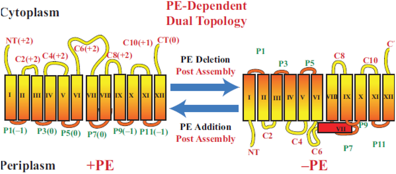
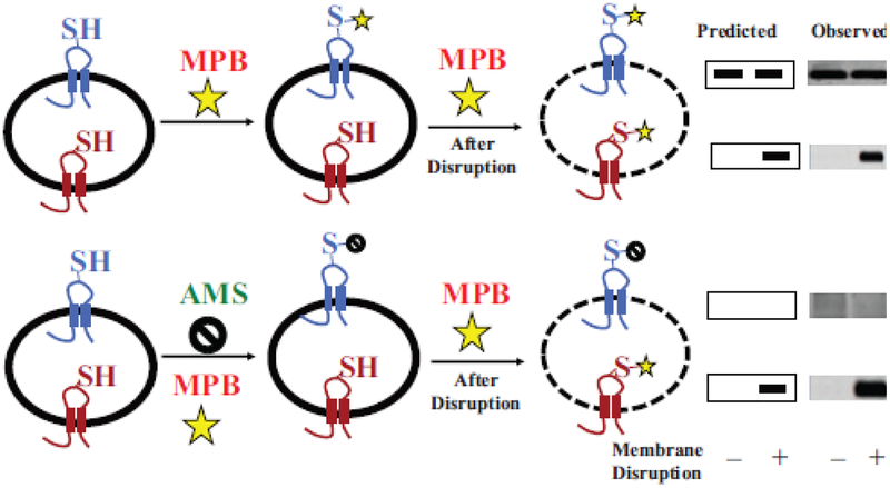
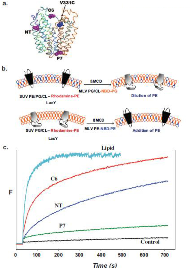
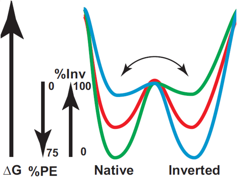
Due to the heterogenous lipid environment in which integral membrane proteins are embedded, they should follow a set of assembly rules, which govern transmembrane protein folding and topogenesis accordingly to a given lipid profile. Recombinant strains of bacteria have been engineered to have different membrane phospholipid compositions by molecular genetic manipulation of endogenous and foreign genes encoding lipid biosynthetic enzymes. Such strains provide a means to investigate the in vivo role of lipids in many different aspects of membrane function, folding and biogenesis. In vitro and in vivo studies established a function of lipids as molecular chaperones and topological determinants specifically assisting folding and topogenesis of membrane proteins. These results led to the extension of the Positive Inside Rule to Charge Balance Rule, which incorporates a role for lipid-protein interactions in determining membrane protein topological organization at the time of initial membrane insertion and dynamically after initial assembly. Membrane protein topogenesis appears to be a thermodynamically driven process in which lipid-protein interactions affect the potency of charged amino acid residues as topological signals. Dual topology for a membrane protein can be established during initial assembly where folding intermediates in multiple topological conformations are in rapid equilibrium (thus separated by a low activation energy), which is determined by the lipid environment. Post-assembly changes in lipid composition or post-translational modifications can trigger a reorganization of protein topology by inducing destabilization and refolding of a membrane protein. The lipid-dependent dynamic nature of membrane protein organization provides a novel means of regulating protein function.

Keywords: Charge Balance Rule; Membrane protein; Phospholipid; Protein folding; Topogenesis.


Conflict of interest statement

Compliance with Ethical Standards: Authors declare no conflicts of interest. No human subjects or animials were used by the authors.

Figures

Figure 1.
Figure 1.
Summary of glycerolipid physical and chemical properties [5]. Glycerolipid headgroups range from net charged (anionic or cationic) to neutral (either uncharged or zwitterionic). R1 and R2 denote acyl chains of fatty acids esterified to diacylglycerol (DAG). Depending on the shape of the molecular when considering the ionized headgroup and the fatty acid composition, these lipids can either be bilayer (cylindrical shaped) or non-bilayer (prism shaped) prone. PE can assume either a cylindrical shape when both fatty acids are fully saturated or a prism shape when at least one fatty acid is unsaturated. CL is non-bilayer in the presence of divalent cations, which is the physiological state. Temperature and fatty acid composition affect both the fluidity (lower with saturated fatty acids and at lower temperatures) and the bilayer to non-bilayer transition, which occurs as temperature is raised. Although cellular membranes are bilayer to maintain barrier function, the presence of non-bilayer prone lipids introduces lateral stress and local disorder within the lipid bilayer.
Figure 2.
Figure 2.
Synthesis of native and foreign lipids in E. coli. The enzymes with their respective genes named catalyze the following steps for synthesis of native phospholipids noted in blue: 1. CDP-diacylglycerol synthase; 2. phosphatidylserine synthase; 3. phosphatidylserine decarboxylase; 4. phosphatidylglycerophosphate synthase; 5. phosphatidylglycerophosphate phosphatases encoded by three genes; 6. cardiolipin synthases encoded by 3 genes; 7. phosphatidylglycerol:pre-membrane derived oligosaccharide (MDO) sn-glycerol-1-P transferase; 8. diacylglycerol kinase. The enzymes with their respective genes named and their source catalyze the following steps for synthesis of phospholipids foreign to E. coli noted in red: 9. phosphatidylcholine synthase (Legionella pneumophila [21,22]); 10. phosphatidylinositol synthase (Saccharomyces cerevisiae [23]); 11. glucosyl diacylglycerol synthase (Acholeplasma laidlawii [24]); 12. diglucosyl diacylglycerol synthase (Acholeplasma laidlawii [25]); 13. lysyl t-RNA:phosphatidylglycerol lysine transferase (Staphylococcus aureus [26]). Figure (modified) and legend reprinted by permission from Springer Nature: [27] Copyright 2018.
Figure 3.
Figure 3.
Topological organization of LacY as a function of membrane lipid composition. TMDs (Roman numerals) and EMDs (Arabic numerals) are sequentially numbered from the N-terminus to C-terminus with EMDs exposed to the periplasm (P) or cytoplasm (C) as in wild type cells. Net charge of EMDs is shown. Topology of LacY is shown after initial assembly in PE-containing cells (+PE) or after initial assembly in PE-lacking (–PE). The interconversion of topological conformers and the ratio of native to inverted conformer are reversible in both directions depending on the dynamic level of PE in membranes. Figure (modified) and legend reprinted by permission from Springer Nature: [27] Copyright 2018.
Figure 4.
Figure 4.
General strategy for SCAM using impermeable thiol reagents. A target membrane protein containing a single cysteine replacement exposed either to the exterior (blue) or interior (red) side of a cell membrane, membrane vesicle or proteoliposome is shown. Half of the sample is treated with the detectable thiol reagent 3-(N-maleimidylpropionyl) biocytin (MPB) to specifically label the externally exposed cysteine (top panel), and the other half is treated with the non-detectable thiol reagent 4-acetamido-4’-maleimidylstilbene-2,2’-disulfonic acid (AMS) (bottom panel) to protect external cysteines during subsequent MPB lableing. Both halves are either kept intact (–) or disrupted (+) by sonication or detergent treatment to expose the interior cysteine followed by treatment with MPB to specifically label the previously inaccessible internal cysteine residue. Labeling by MPB that can be blocked completely by pretreatment with AMS is an independent verification of an outside facing residue (bottom panel). Labeling by MPB that cannot be blocked by such AMS treatment is an independent verification of a residue that is facing the cytoplasm. The target protein is immunoprecipitated and resolved by sodium dodecyl sulfate-polyacrylamine gel electrophoresis and biotinylated protein is detected using avidin-horse radish peroxidase with the predicted and observed results shown on the right. A protein with dual topology would display an increase in the amount of labeled protein after disruption in the upper panel and reduced amount of labeling detected after disruption in the lower panel. The modified figure and legend were reprinted by permission from Springer Nature: [49] Copyright 2019.
Figure 5.
Figure 5.
Monitoring lipid-dependent topological changes in fliposomes. a. Schematic native structure of LacY showing the position of an engineered tryptophan residue in either EMD NT (residue 14), C6 (residue 205) or P7 (residue 250) relative to the chromophore at position V331C. b. LacY engineered to display high FRET intensity in the native conformation (upper left) or low FRET intensity in the inverted conformation (lower left) was reconstituted into small unilamellar vesicles (SUV) with or without PE, respectively. The SUVs contained trace amounts rhodamine labeled PE. Multilamellar vesicles (MLV) containing PG and CL with a trace amount of 6-[(7-nitro-2–1,3-benzoxadiazol-4-yl)amino]hexanoyl-PG (NBD-PG) or PE with a trace amount of NBD-PE were added to the SUVs containing native or inverted LacY, respectively. Transfer of lipids between SUVs and MLVs was initiated by addition of ß-methyl cyclodextrin (ßMCD)-loaded MLVs to the SUV suspension. The rate of lipid transfer was monitored by FRET between rhodamine- and NBD-labeled lipids. The rate of LacY topological change was monitored by FRET between a tryptophan residue and a chromophore in the C-terminal six TMD bundle of LacY. c. Time scale for change in FRET upon addition of PE to proteoliposomes containing LacY in the absence of PE. Control indicates lack of changes in FRET when SUVs and MLVs both lack PE. Figure 5b was reproduced from[98] by permission of the National Academy of Sciences, USA. Figure 5c was constructed based on data presented in [98].
Figure 6.
Figure 6.
Dual minima energy folding funnel for LacY as a function of membrane lipid composition. The folding of LacY to its lowest free energy state (ΔG) proceeds via a funnel-shaped energy landscape whose shape is defined by the physicochemical properties of the lipid environment (green, 75% PE; red, intermediate % PE; blue, 0% PE). The conformational space available to the population of folding protein conformers at a given lipid composition is defined by the funnel circumference (x-axis) and the internal ΔG (y-axis) of each folding intermediate. As LacY folds to lower energy conformations, it populates thermodynamic traps whose depth and shape determine the percent of the final native or inverted conformation at steady state. Membrane lipid composition affects a late folding event, which is postulated here to define a rapid equilibrium (horizontal arrow) between subsequent pathways leading to either the native, inverted or mixed conformation separated by a high thermodynamic barrier. Figure and legend reproduced from [50] by permission of American Society for Biochemistry and Molecular Biology

References

    1. Blobel G (1980) Intracellular protein topogenesis. Proc Natl Acad Sci U S A 77:1496–1500. - PMC - PubMed
    1. Kuhn A, Koch HG, Dalbey RE (2017) Targeting and Insertion of Membrane Proteins. EcoSal Plus 7. - PMC - PubMed
    1. Rapoport TA (2007) Protein translocation across the eukaryotic endoplasmic reticulum and bacterial plasma membranes. Nature 450:663–669. - PubMed
    1. Cymer F, von Heijne G, White SH (2015) Mechanisms of integral membrane protein insertion and folding. J Mol Biol 427:999–1022. - PMC - PubMed
    1. Dowhan W (1997) Molecular basis for membrane phospholipid diversity: why are there so many lipids? Annu Rev Biochem 66:199–232. - PubMed

Publication types

MeSH terms

LinkOut - more resources