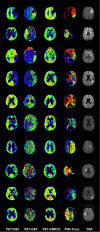
Penumbra detection in acute stroke with perfusion magnetic resonance imaging: Validation with 15 O-positron emission tomography
- PMID: 30937950
- PMCID: PMC6593670
- DOI: 10.1002/ana.25479
Penumbra detection in acute stroke with perfusion magnetic resonance imaging: Validation with 15 O-positron emission tomography
Abstract
Objective: Accurate identification of the ischemic penumbra, the therapeutic target in acute clinical stroke, is of critical importance to identify patients who might benefit from reperfusion therapies beyond the established time windows. Therefore, we aimed to validate magnetic resonance imaging (MRI) mismatch-based penumbra detection against full quantitative positron emission tomography (15 O-PET), the gold standard for penumbra detection in acute ischemic stroke.
Methods: Ten patients (group A) with acute and subacute ischemic stroke underwent perfusion-weighted (PW)/diffusion-weighted MRI and consecutive full quantitative 15 O-PET within 48 hours of stroke onset. Penumbra as defined by 15 O-PET cerebral blood flow (CBF), oxygen extraction fraction, and oxygen metabolism was used to validate a wide range of established PW measures (eg, time-to-maximum [Tmax]) to optimize penumbral tissue detection. Validation was carried out using a voxel-based receiver-operating-characteristic curve analysis. The same validation based on penumbra as defined by quantitative 15 O-PET CBF was performed for comparative reasons in 23 patients measured within 48 hours of stroke onset (group B).
Results: The PW map Tmax (area-under-the-curve = 0.88) performed best in detecting penumbral tissue up to 48 hours after stroke onset. The optimal threshold to discriminate penumbra from oligemia was Tmax >5.6 seconds with a sensitivity and specificity of >80%.
Interpretation: The performance of the best PW measure Tmax to detect the upper penumbral flow threshold in ischemic stroke is excellent. Tmax >5.6 seconds-based penumbra detection is reliable to guide treatment decisions up to 48 hours after stroke onset and might help to expand reperfusion treatment beyond the current time windows. ANN NEUROL 2019;85:875-886.
© 2019 The Authors. Annals of Neurology published by Wiley Periodicals, Inc. on behalf of American Neurological Association.
Conflict of interest statement
Nothing to report.
Figures


References
-
- Astrup J, Siesjo BK, Symon L. Thresholds in cerebral ischemia — the ischemic penumbra. Stroke 1981;12:723–725. - PubMed
-
- Baron JC. Mapping the ischaemic penumbra with PET: implications for acute stroke treatment. Cerebrovasc Dis 1999;9:193–201. - PubMed
-
- Heiss WD. Ischemic penumbra: evidence from functional imaging in man. J Cereb Blood Flow Metab 2000;20:1276–1293. - PubMed
-
- Heiss WD, Huber M, Fink GR, et al. Progressive derangement of periinfarct viable tissue in ischemic stroke. J Cereb Blood Flow Metab 1992;12:193–203. - PubMed
-
- Hossmann KA. Viability thresholds and the penumbra of focal ischemia. Ann Neurol 1994;36:557–565. - PubMed
Publication types
MeSH terms
Substances
Grants and funding
LinkOut - more resources
Full Text Sources
Medical

